Potential occupational, non-occupational and accidental radiation exposures in industrial radiography using radioactive sources
Published: Dec 12, 2023
A quick word from Ionactive
If you like this blog article and need formal radiation safety training, such as our online multimedia 24/7 RPS course, then please get in touch. Other radiation protection courses are available (including onsite face to face bespoke training). This free resource scrapes the surface of the type of training material we cover (this material within our training courses is presented in videos, audio commentary, slide shows, interactive 'widgets', animations, text, quizzes and much more).
Industrial Radiography
We spent some time discussing what industrial radiography is in this recent blog article: 'When is an 'industrial radiography walk-in enclosure' not an industrial radiography enclosure (and therefore does not require an HSE consent)?'. You may want to pop briefly to this article, or just continue with our summary below.
Industrial radiography is the use of ionising radiation in non destructive testing (NDT). NDT is generally carried out on industrial pipe welds, safety critical structures such as pressure vessels or jet engine turbine blades and similar. If industrial radiography is taking place in Great Britain then this requires a consent from HSE, or HSENI in the case of Northern Ireland. If the work is taking place on a nuclear licensed site then the consent is obtained from ONR. We have provided significant comment and guidance on the consent process at the following blog link: 'New UK Consent process for users of Ionising Radiation' (article updated October 2023).
Where the work is carried out in an enclosure (e.g. x-ray cabinet) which cannot be entered (i.e. cannot be walked into), then it can still be classed as NDT (if it is NDT), but only a registration is required - this is then NOT classed as industrial radiography.
Where industrial radiography is taking place, then it can take one of two forms which are summarised below.
Enclosure Industrial Radiography
Where the work is carried out in a purpose made enclosure (that can be walked into), with appropriate safety systems, interlocks, active signage, fail safe interlocks (etc), it is known as enclosure radiography and this is always the preferred option (the ALARP option). Where an item can be reasonably carried / transported, then enclosure radiography should take place.
Site Industrial Radiography
Where using a purpose made enclosure is not reasonably practicable then site industrial radiography may have to take place. With this technique you take the source of ionising radiation (radioactive source / x-ray system) to the item requiring NDT. It follows that this item is generally going to be large and / or fixed in position where it cannot be easily moved to an enclosure. This type of industrial radiography is assumed to have higher exposure risks and relies predominantly on maximising distance, and using local shielding and collimation to limit direct beam or scatter exposures. The area will be barriered off with passive signage, and there may be flashing lights / audible alarms to warn those in the vicinity that a "shot" (radiographic exposure) is about to take place / is taking place. There is significant reliance on human factors (supervision, management, following correct procedures, security and observation of the area, other persons following signage and similar).
Consent for Industrial Radiography
The consent for industrial radiography is the same regardless of the methods noted above. Where site radiography takes place the radiographic contactor will need to receive 7 days notice of this work in order to allow both contractor and client time to review the work, develop risk assessments and method statements and put in place appropriate mitigations. In exceptional cases, where 7 days notice cannot be given, the client can apply for an emergency waiver from HSE.
What does the consent say about the source of ionising radiation used? Nothing! Industrial radiography is a specified practice and it is the work (NDT) that matters, not the type of source used. Types of ionising radiation sources that could be used include:
- Radiation generators (x-ray generator, neutron generator, betatron, linear accelerator etc).
- Radioactive sources (e.g. Co-60, Ir-192, Se-75).
Note there is a separate consent for operation of an accelerator, but this does not apply in this case since the work is industrial radiography (and the work matters, not the source of ionising, as already stated). Likewise, there is a consent for HASS sources, but this does not apply to industrial radiography since it has its on specific consent.
This has hopefully provided a basic overview of industrial radiography definition and application. The rest of this blog article will now consider the types of facilities and equipment used, before considering the radioactive sources available and typical dose rates during occupational and non-occupational exposures, and in reasonably foreseeable accidents. At all points the radiation safety features of industrial radiography will be described.
Radioactivity (correct units of activity vs tradition)
In the UK (and most of the world) the unit of activity is the becquerel (Bq). All formal records, transport documentation, labels, risk assessments (etc) must refer to the Bq. However, industrial radiography goes back a long way, to a time where the unit of activity was the curie (Ci) - this is still used in the USA. Radioactive sources for purchase are still sold in convenient curie quantities (such as 10 Ci, 20 Ci, 50 Ci etc. These activities translate into the SI system (Bq) as follows:
- 10 Ci = 370 GBq
- 20 Ci = 740 GBq
- 50 Ci = 1.85 TBq
We mention this because even today veteran and more recent industrial radiography technicians in the UK will still refer to their 10 Ci source. In fact, this practice is often referred to as 'bombing' and you may hear the expression '10 Ci bomb'!
General radiation safety aspects of using radioactive sources in industrial radiography
We are not experts in the practice of NDT (industrial radiography) so will not discuss contrast sensitivity, focus-to-film distance (ffd), object-to-film distance or similar. In this section we will discuss some general aspects of radioactive source use in NDT by first considering the projector.
Use of a projector
Generally a projector is used to store the radioactive source for transport and as a means of remotely deploying the source to make an exposure and then retracted back safely for the next exposure (also known as a "shot"). Whilst some projectors are designed for transport within their own right [e.g. Type B (U) Certification / Type A Approval (IAEA TS-R-1) ], others are designed so they have an outer shielding overpack which must be used to achieve designed transport compliance.
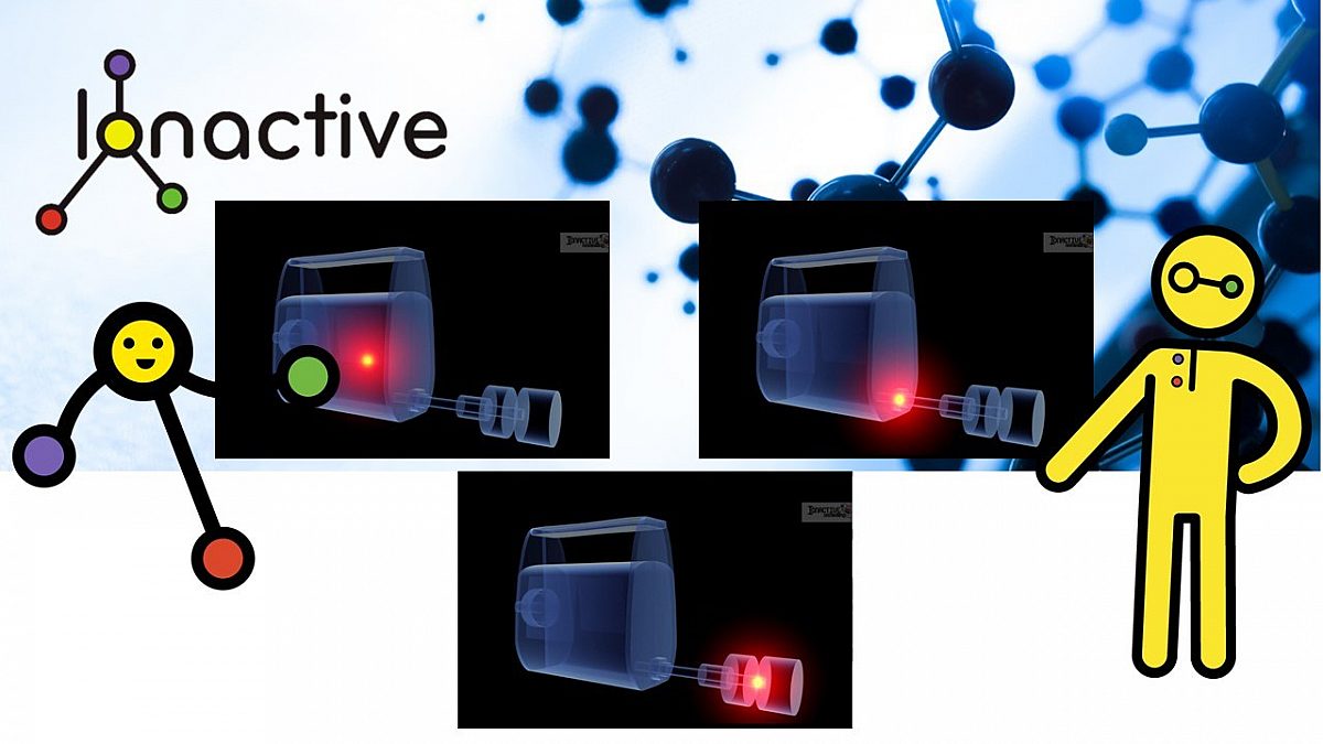
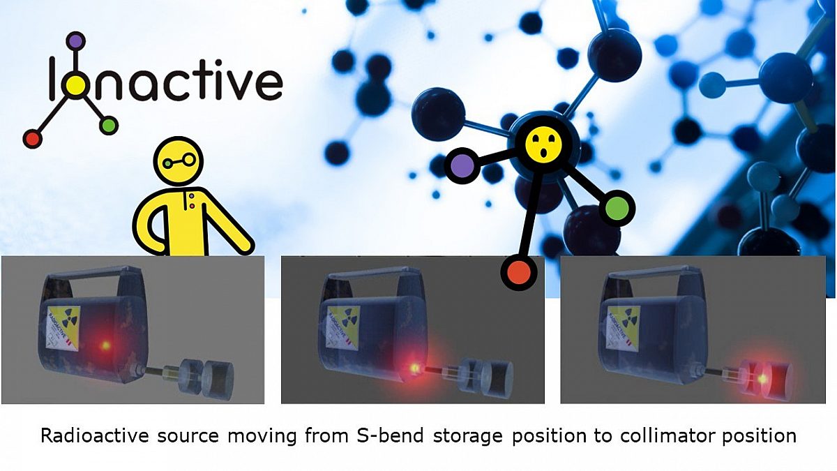
The projector will contain a tungsten (or depleted uranium) "S-bend" source shield. When the source is safely stored it will be in the centre of the bend (so no 'line of sight' out of the projector). Upon attaching a source guide tube to the front of the projector, and a source drive cable to the back, it is possible using a handle (or motor) to drive (push) the source out of the projector, and then retract (pull) the source at the end of the exposure.
Mechanical safety locks ensure that the drive cable has to be securely attached to the projector before the source can be exposed.
Whilst details will vary between projector models (and their age), the back end of the unit will generally have the following features:
- An unlock / lock key or equivalent.
- Turning to 'unlock' allows the source drive cable to interface with the projector.
- The drive cable can then be securely connected to the back end of the source cable. The radioactive source is permanently attached to a small length of drive cable (known as a pigtail) which rests within the S-bend, with its connector (pigtail connector) visible at the back of the projector when the security cover is removed. The longer drive cable connects up to the source when attached to the projector.
- Mechanical safety features ensure that the connection between the shorter pigtail and longer drive cable is secure before the source can leave the projector.
For some industrial radiography shots, such as panoramic radiography, the source guide cable at the front of the projector is replaced with a panoramic collimator. In this configuration the whole projector is placed into position (e.g. inside a large steel pipe), and the source is then remotely driven out into the collimator.
The following graphic presents some of the detail just discussed.
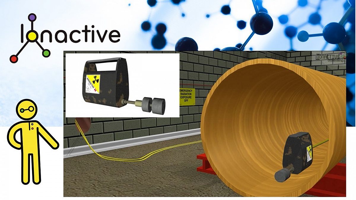
Industrial radiography projector fitted with a panoramic collimator in a steel pipe
In the graphic above we are showing a panoramic collimator fitted onto a projector which has been placed inside a wellhead pipe. You can just see the drive cable leaving the back of the projector, this has been run through the wellhead, and you see it again disappearing into an exit hole in the wall. This graphic therefore depicts an example of enclosure radiography. We will show further features of this enclosure later in the blog.
Below we see a representation of the source moving in the projector from storage position, around the S-bend and out into the collimator.
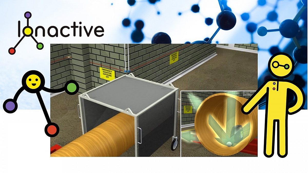
In the next graphic we see a representation of the radiation streaming from the source through the collimator and onwards through the pipe. We have already mentioned that this is in an enclosure, so have a think about where the diagonal gamma ray path might be heading.

Gamma ray paths from the source, through the collimator and onwards though the pipe
Industrial Radiography Enclosure
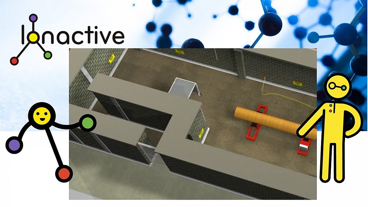
Let's now reveal the enclosure we were talking about earlier. The first view is from the side.
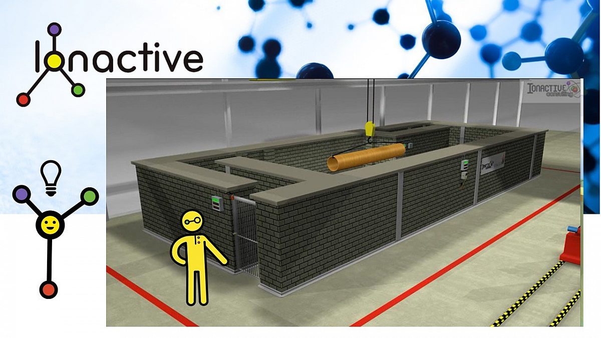
Side view of the industrial radiography enclosure
And below is a top view of the enclosure. At this point you may already be able to note three possible radiation exposures from this setup.
- Occupational exposure - potential sky-shine dose rates at ground level caused by scattered radiation above the enclosure --> impact on the radiographer.
- Non-Occupational exposure - potential sky-shine dose rates at ground level caused by scattered radiation above the enclosure --> impact on other employees working in the area.
- Accidental exposure - an unplanned exposure to a person working outside the enclosure.
We will also consider direct occupational exposure of radiographers working inside the enclosure.
Sky-shine is something we will also consider later, but follow the link if you want to review this phenomenon right now.

Top view of the industrial radiography exposure
Ideally this enclosure would have a shielded roof. However, there are some circumstances where this is not possible when moving massive objects in and out. With calculation, local shielding, and suitable workflow, dose rates outside the enclosure can be shown to be ALARP, and in any case below the level of a controlled area. Despite not having a shielded roof, this set up is much better then relying on open site radiography.
Open Site Industrial Radiography
Open site radiography could be represented as shown in the following graphic.

Representation of open site industrial radiography
The projector equipment represented above is little different to that already discussed. The main difference is that with site radiography the source tends to be deployed by manual means using a handle (hand crank) as shown, whereas with the enclosure radiography the source deployment is driven by an electric motor which is interlocked into the enclosure safety systems (more of that in a moment).
Site radiography features include:
- Demarcation of the area by a barrier.
- Passive warning signs placed around the perimeter (Controlled Area - Gamma sources / Keep Out etc).
- Radiation protection mostly provided by maximising distance from the source.
- Radiation protection further enhanced by collimation of the source and local shielding (and possibly also the intrinsic shielding provided by the radiographic object).
- Visible active warning signs (e.g. strobe lights).
- Audible indicators (e.g. audible warning before source is about to be deployed).
- Real time radiation monitoring of the perimeter by radiography technicians (as a minimum looking for < 7.5 micro Sv/h instantaneous dose rate).
All the above protective features rely on human factors - suitably qualified and experienced persons doing the right thing. This is why enclosure industrial radiography is preferred (as safety is enhanced with engineered safety systems). For this reason we will now consider some of the engineered safety features of enclosure radiography which make it so much more preferable.
Enclosure radiography - engineered features for radiation protection
We will continue to examine our featured enclosure, other enclosures may be different (and many will have a shielded roof which is always preferable if reasonably practicable).
If you go back and look at the two views of our enclosure you will note the following engineered features for radiation protection. A slide show of the following descriptions follows below.
- Shielding walls to reduce direct radiation dose rate to < 1 micro Sv/h (whilst < 7.5 micro Sv/h might be in IRR17, the ALARP principle can be applied to lower that still further).
- (No need for) barrier tape and temporary warning signs or restriction of employees to large areas of the site.
- Open top enclosure - for our specific example it is not practical to shield the roof. This therefore provides some interesting radiation protection challenges due to sky-shine (scattered radiation above the enclosure).
- Entry via a labyrinth (maze) which lowers radiation dose rates to background levels at the gates (no shielding door is required). In our example the enclosure has two points of entry / exit.
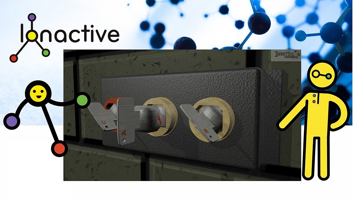
- Trapped castell key gate locks (when gate is unlocked and opened this key is trapped).
- Gate interlock (secondary safety device, whereas the key is the primary device). Should the gate lock fail, opening the gate would sound an alarm and retract the source (details to follow). This is known as defence in depth since two independent coincident failures would need to occur before an open gate could result in an unplanned exposure.
- Emergency pull cables inside the enclosure - pulling these will either 1) stop the source deployment if in the imminent stage, or 2) cause retraction of the source if exposure is already taking place.
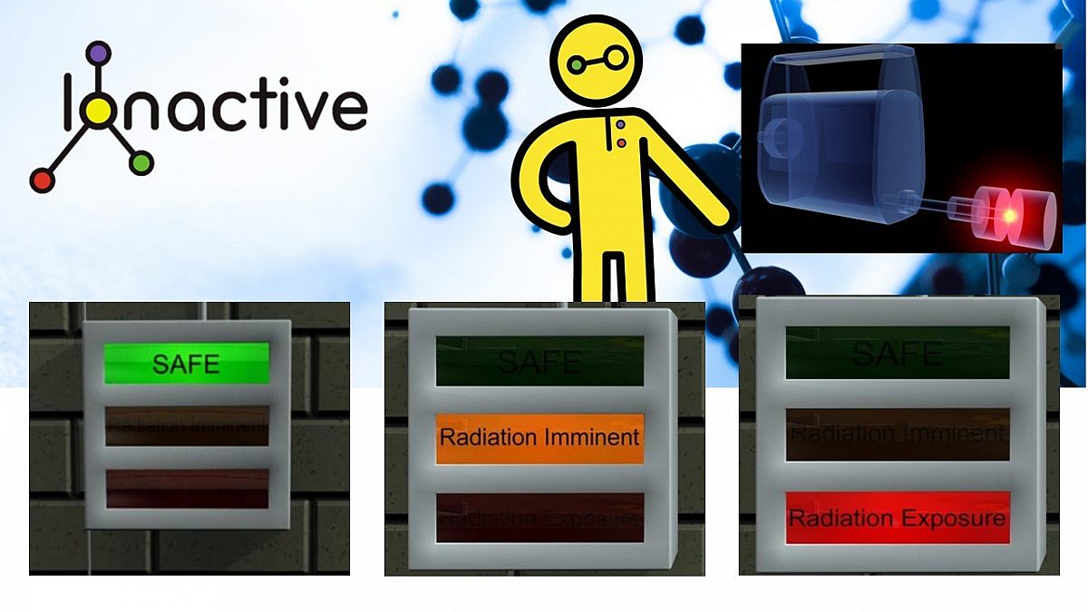
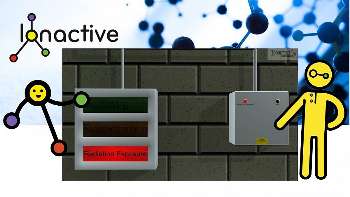
- Active, 3 stage warning light boxes (two inside the enclosure and four mounted on the walls outside the enclosure). These show "safe" (green) meaning system on and safe, "Radiation imminent" (amber) meaning that the radioactive source is about to be deployed, and "Radiation exposure" (red) meaning that the source is deployed (outside the projector). In this facility the amber and red lights "fail safe" so a failure means the source cannot deployed (or will be retracted upon failure if already deployed).
- Independent radiation monitor - mounted inside the enclosure. If the radioactive source is not safely in the projector (for whatever reason) and despite other safety system indicators, this monitor detects radiation above background and will ensure that the red "Radiation exposure" indicator remains illuminated. The threshold is set so that a fully loaded projector with source safely stored does not set off the alarm (the exposure indicator will only illuminate which significant levels of exposure are detected above background).
- The presence of the source projector in the bay (with connections made) will ensure, as a minimum, that the green "safe" light is illuminated, indicating that the radiography exposure system is live. If the projector is disconnected the safe light will not be illuminated (indicating source deployment system is switched off).
- Use of local collimation and shielding. In our example collimation is present via the panoramic collimator. In addition, a mobile local shield is also deployed to minimise radiation scatter (sky-shine) above the enclosure (see pictures below). This is an example of a human factor derived protective feature working with the engineered safety features.
- When the castell key is removed from the gates (A) and (B) (i.e. locked shut) the gate interlock previously mentioned detects this closure (i.e. additional confirmation of closure).
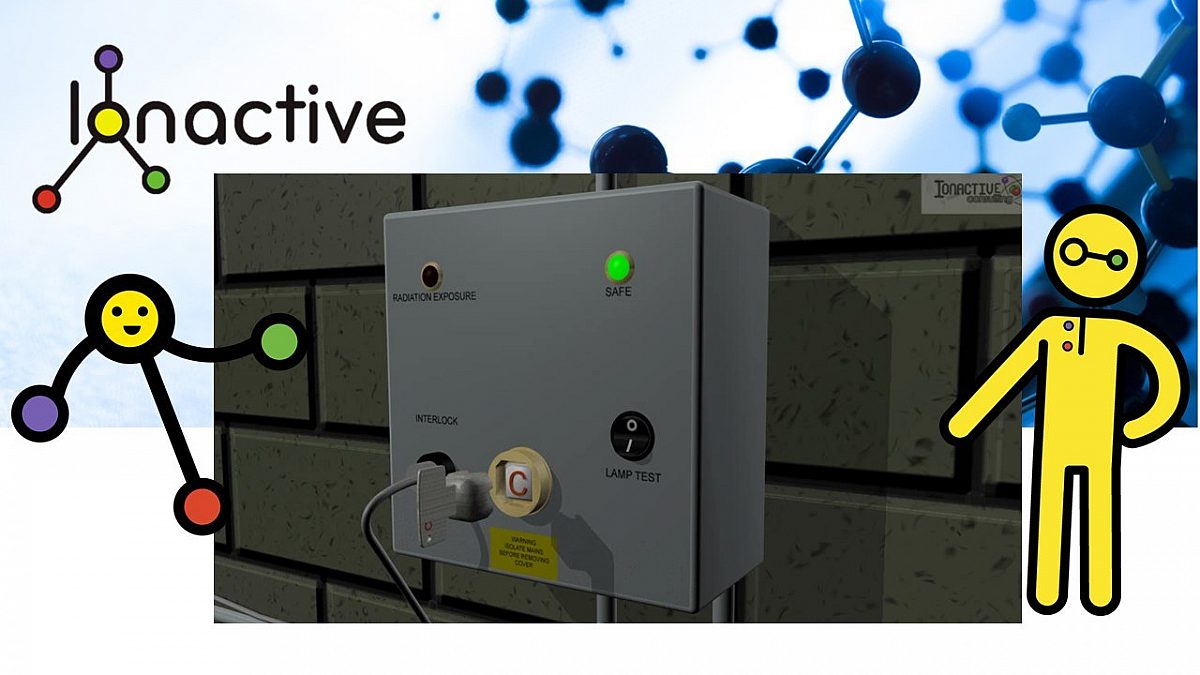
- Castell key exchange box (near control panel). This ensures that both gates to the enclosure are shut (i.e. keys removed and placed into the exchange box). Once both keys are in the exchange box and locked into position, a third (red) key (C) is released which is placed into the interlock control box.
- Interlock control box. This powers the source motor, monitors the status of active signage (including failure), monitors gate interlocks, the independent radiation monitor and emergency pull cables. When this third key (C) is used to energise the radiography system, the two gate castell keys (A & B) remain locked into the exchange box.
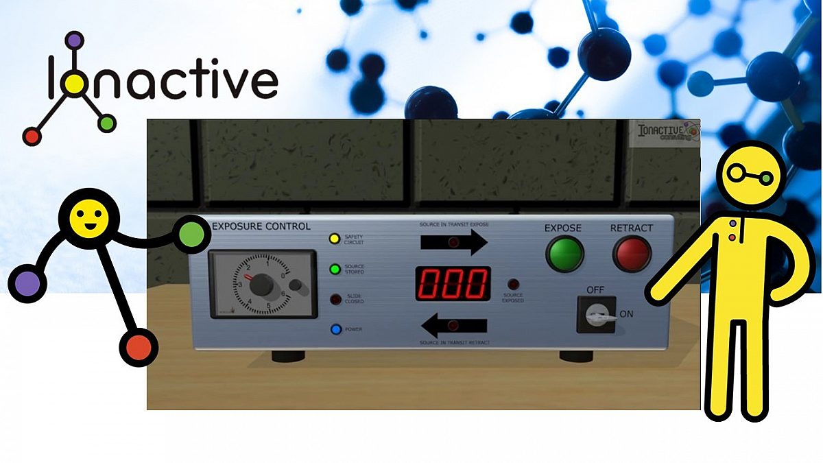
- Exposure control box. This is connected to the interlock control box which is used by the radiography technician to initiate and terminate radiation exposures. The box includes safety status lights (e.g. yellow safety circuit indicator, green source storage indicator), source in transit indicator (for deployment and retraction), a timer (for auto exposures), a green expose button and a red retract button. This box also has an on / off key switch.
- If the green exposure button is pressed (and all safety systems are set correctly - i.e. yellow indicator), then an audible alarm will sound for 5 seconds inside / outside the enclosure and an amber radiation imminent message will be illuminated inside and outside the enclosure.
- Source wind-out motor (powered by interlock control box and operated from the exposure control box). This is mounted outside the enclosure, and the source drive cable attaches to (and from) the motor via a small duct in the shielding wall (the duct is sloped through the wall so there is no direct line of sight into the enclosure). If there was a power failure a handle can be attached to the motor drive to manually manipulate the source (this handle is locked away and only used in an emergency to avoid unauthorised movement of the source).
A search and lock up system is potentially missing from this system. This would require a LPO (last person out) button to be pressed inside the enclosure which would initiated a timed sequence (with audible indicator). There would be perhaps 20 seconds to vacate the enclosure and shut the gate(s) before pressing a LPC (last person confirm) button (on the outside wall near the gate). If this sequence is not followed exactly in the time allowed, the whole process would need to be started again. For very large enclosures with lots of hidden space there may be more than one LPO button, each needing to be pressed in sequence to ensure the whole area is checked. In the example given in this blog it was decided that a LPO / LPC system was not required.
Some of the features above are now presented in the slide show below.
This run through of the safety features of this enclosure demonstrates why this is preferred to open site radiography.
We will now move away from describing the radiography equipment and take a look at the radiation emission properties of typical industrial radiography sources.
Radioactive sources used in industrial radiography
There are three main radionuclides used in industrial radiography - in descending order of energy they are:
- Cobolt-60 (Co-60)
- Iridium-192 (Ir-192)
- Selenium-75 (Se-75)
Co-60 and Ir-192 are beta / gamma emitters, whereas Se-75 decays by electron capture, yielding gamma rays. In all cases it is the gamma rays which are important when performing industrial radiography.
The choice of what to use is outside the scope of this blog and into the realms of NDT, but we can provide a few considerations here. Generally, the thicker & denser the material and / or the shorter exposure time required, you would choose a higher energy emitter. This is not quite true in all cases since a relatively thin lower density material, with a high energy gamma emitter, might not produce the contrast required. However, what is true is that for a given activity (say 1 GBq) and distance (say 1 m), the dose rate will be higher as the gamma energy increases. It is also true that as the energy increases the 10th value thickness (TVT) for a given material (e.g. steel) will also increase.
To be compatible with the principles of radiation protection, the minimum activity and gamma energy emitter should be chosen which still meets the radiographic (NDT) requirements of the work. However, this is not always achieved as highlighted in the following examples:
- The time of each 'shot' will be influenced by activity, so putting NDT work (especially open site radiography) between other work may lead to higher activities being used.
- Particularly for Ir-192 with a relatively short half life (74 days), there is a temptation to purchase sources of higher activity (and therefore dose rate output), to make best use of the source over the following couple of months.
All three radioactive sources, at the activities used in industrial radiography, will be classed as HASS (High Activity Sealed Sources). In the UK this will mean that users will need a HASS permit from the relevant environment agency of the devolved administrations. In turn this will require a significant security regime which will be inspected by the relevant agency and the police via the CTSA (Counter Terrorism Security Adviser). In terms of health and safety under the Ionising Radiations Regulations 2017 (IRR17), use of the sources will require a consent from HSE. Since the specified practice is 'Industrial Radiography' it is for this practice where the consent is held, and not for HASS sources (use of HASS sources which does not include industrial radiography or industrial irradiation has their own consent category).
All this leads us to state that industrial radiography radioactive sources can be considered highly dangerous and must be careful controlled by suitably qualified persons under the permitting / consent regime. X-ray NDT is not the subject of this blog as stated at the start, but it is clear there is a preference for x-ray where reasonably practicable - since this removes the security challenges of HASS sources since the radiation hazard will cease the moment power is removed. The dose rate output of high kV / mA x-ray tubes can also be highly dangerous too, but radiation protection is still much simplified. Reasons where x-ray tubes may not be suitable include:
- The energy (kV) or rate of emission (mA) [both of which combine to give dose rate] may not be high enough for the NDT image required.
- X-ray tubes tend to be heavy and bulky, and when combined with the heavy duty HT cable (which may also contain cooling air / water), may not fit into the space required to make the shot.
- The location may not have a suitable power supply for the x-ray generator (more likely for open site radiography).
- Required collimation and minimum / maximum focal size may be more difficult to achieve with x-ray (noting the physical size of the radioactive source is tiny compared to an x-ray head). Typical dimensions of an Ir-192 source used in NDT will be about 6mm diameter and up to 24mm in length (this is for the outer capsule).
Radiation protection related properties of radioactive sources used in industrial radiography
Find below basic radiation protection related properties for each of the three radioactive sources featured above. This section of the blog will provide data which we can then use to describe likely occupational and accidental exposures.
Cobolt-60 (Co-60)
- Type of emission - Beta / Gamma
- Gamma emission energy - 1.17 and 1.33 MeV (100%)
- Half life - 5.3 years
- Dose rate in air unshielded at 1m per GBq - 0.306 mSv/h
- Dose rate in air unshielded at 1m [typical source of 100 Ci / 3.7TBq] - 1135 mSv/h
- TVT Steel - 73mm (HVL - 22mm)
Iridium-192 (Ir-192)
- Type of emission - Beta / Gamma
- Gamma emission energy - 0.317 MeV (83%), 0.468 MeV (48%), 0.604 (8%)
- Half life - 74 days
- Dose rate in air unshielded at 1m per GBq - 0.113 mSv/h
- Dose rate in air unshielded at 1m [typical source of 15 Ci / 555 GBq] - 62.8 mSv/h
- TVT Steel - 43 mm (HVL - 13 mm)
Selenium-75 (Se-75)
- Type of emission - electron capture (-> gamma)
- Gamma emission energy - 0.136 MeV (59%), 0.265 MeV (59%), 0.401 MeV (12%)
- Half life - 120 days
- Dose rate in air unshielded at 1m per GBq - 0.046 mSv/h
- Dose rate in air unshielded at 1m [typical source of 15 Ci / 555 GBq] - 25.4 mSv/h
- TVT Steel - 27 mm (HVL - 8 mm)
If required used our glossary for some of the terms above such as TVT. This concept is also explained nicely here: How do I convert TVT to HVT (or the other way around)?
Occupational exposure scenarios
In this section we will describe several occupational exposure scenarios. Some are based on generic assumptions and general data, others are more related to the radiographic equipment and sources we have highlighted thus far.
Occupational exposures from handling projectors
Anyone who has worked with a projector will know that handing duration is minimised by the fact they are heavy and have manual handling risks. Once the projector has been moved into position (e.g. from storage location / transport van), the ALARP principle requires that exposures are minimised by maximising the distance between the operator and projector.
The maximum dose rate from the surface of a typical radiography projector will be < 2 mSv/h (depending on package category). A transport index (TI) of 0.1 will infer a dose rate at 1m from the projector of 1 micro Sv/h, and an index of 1.0 will infer a dose rate at 1m of 10 micro Sv/h. Both these TI values would mean the projector was a category II-yellow and that the surface dose rate was between 5 micro Sv/h and less than 500 micro Sv/h.
A typical example of a projector might be 10 Ci (370 GBq) of Ir-192 and in our example below has a transport index of 0.1 (1 micro Sv/h at 1m).

370 GBq Ir-192. TI of 0.1 is 1 micro Sv/h at 1m & < 0.5 mSv/h on surface
For most of the time it is reasonable to assume that the NDT technician is at least 1m from the projector, and at 1 micro Sv/h implies occupational exposure around the projector will not be significantly above background.
There will be times where the projector is carried from a vehicle to the the work area. Given the mass of the protector, this will not be a long task - let as assume 1 minute of manual handling per task, 5 days per week, 50 weeks per year (by the same technician - assumed to be a critical person). Whilst the inverse square law will not work perfectly with such a 'large' source (projector), we can use it to infer a whole body dose rate which might be located some 10cm from the projector. This is simply 1002 / 102 x 1 micro Sv/h which yields 100 micro Sv/h.
This could present an occupational whole body exposure of 417 micro Sv/year. There is also the work required around the projector when connecting up the source drive cable etc, however since this is now much less of a manual handling issue, using ALARP to maximise distance (i.e. working at arms length) is reasonably practicable (so dose rate probably tends nearer to the 1 micro Sv/h).
Whilst carrying the projector there is also a potential extremity dose to the fingers. In our example we might assume the surface dose rate is up to 500 micro Sv/h. Whilst the handle is a few cm's away from the surface, 500 micro Sv/h seems a usable upper level for our example.
[Ionactive comment. We note that for a II-yellow projector, the TI can vary from 0.1-1 and the surface dose rate will vary from 5 micro Sv/h up to just less than 500 micro Sv/h. This will imply that with a TI of 0.1 the surface dose rate will be nearer 5 micro Sv/h because each increase in TI (i.e. 0.1-0.2, 0.2-0.3) would linearly increase the dose rate by just less than 50 micro Sv/h each time. However, our analysis is not based on real measurements so it is easier to use the maximums in each category (therefore we choose a surface dose rate of 500 micro Sv/h regardless of TI between 0.1-1].
Using the same analysis above we find a potential dose uptake of about 2mSv/year to the extremities. Not huge compared to the legal limit of 500 mSv/year equivalent dose to the extremities (and noting the classified person threshold of 150 mSv/year).
[Ionactive comment: We do wonder what percentage of industrial radiography technicians would routinely use extremity dosimetry whilst carrying the projector?].
This example is based on a relatively modest 370 GBq (10 Ci) of Ir-192 so the dose rates near the projector are likely to be less than specified above. Recall that the projector might be rated up to say 5.55 TBq (150 Ci) of Ir-192 and the higher activities could result in a TI of between 1 and 10 (suggesting up to100 micro Sv/h at 1m and up to just less than 2 mSv/h on the surface). Ionactive experience shows these higher activities are generally less common and / or take place in an enclosure where there may be less manual handling of the projector. It would therefore not be appropriate to simply factor up our estimates based on increased activity.
We will now consider some site industrial radiography based on our 370 GBq Ir-192 projector initially, and then explore different activities and radionuclides.
Occupational exposures from using projectors in site radiography
In the discussion which follows we will assume the projector is already in place and ready to be used - occupational exposure estimates to get to this point (transport and attaching drive cables etc) were discussed above.
The graphic below reminds us of what we are dealing with: an open area barriered off with tape, passive warning signs, supervision by eye, using audible and visual warning devices (e.g. sounder and strobe light) etc.

Representation of open site industrial radiography
We will firstly consider dose rates from 370 GBq of Ir-192 in air (i.e. no collimator or shielding from the radiographic object).
We need to ensure that no controlled area exists outside of the perimeter. IRR17 ACoP para 298 specifically provides guidance that for industrial radiography a controlled area will exist where the dose rate exceeds 7.5 micro Sv/h when averaged over a minute. So the 'line in the sand' is 7.5 micro Sv/h, but the ALARP principle would be to aim for less where reasonably practicable.
Have a look at the following graphic and consider the position of the radioactive source in transit (red dot) between the projector and the collimator (attached to the pipe).

Radioactive source in transit between the projector and the radiographic object
Note that the distance A (between bottom barrier and source in transit) is LESS than distance B (between bottom barrier and radiographic object). Also note that at point C the source is partially shielded by a combination of the radiographic object and a collimator (which is directing the radiation down onto / into the pipe). During the transit of the source from projector to collimator it is unshielded and is nearer the bottom barrier. Therefore, the instantaneous dose rate (IDR) during source transit will be higher at the bottom barrier than once the source is deployed. This is mostly ignored since the transit time is a couple of seconds at most (and significantly less than the dose rate averaged over one minute).
But keep something in mind (and this is specifically explored later under accidents / incidents). What would the dose rate be at the bottom barrier if the source becomes stuck in the guide tube at the position shown?
Distance B - from source to bottom barrier (no collimator etc)
Using the radioactive source data from earlier we note that the dose rate 1m from the unshielded 370 GBq Ir-192 source will be 41.82 mSv/h. We need this down to (less than) 7.5 micro Sv/h as noted above. Using the inverse square law we can state the following:
\[\frac{D1}{d_{2}^{2}}=\frac{D2}{d_{1}^{^{^{2}}}}\]
D1 is the dose rate at distance d1 and D2 is the dose rate at distance d2. So we can say that D1 is our dose rate at 1m (d1) and we need to find the distance (d2) which brings the dose rate D2 down to 7.5 micro Sv/h (or less). We we can rearrange:
\[d_{2}=\sqrt{\frac{D_{1}d_{1}^{2}}{D_{2}}}\]
We then can pop the numbers in where all the dose rates are in micro Sv/h and the distances are in m as shown below:
\[d_{2}=\sqrt{\frac{41820\times(1)^{2}}{7.5}}\]
This shows that d2 needs to be at least 74.7m - which is the distance (B) between the exposed source and barrier in the graphic above. This is a slight exaggeration as there would be photon air attenuation over this distance (probably bringing distance down to somewhere near 66m). In most site radiography applications this is just not reasonably practicable - if there are persons located around the perimeter in all directions then you have an exclusion zone of the order of 5400 m2.
Clearly something has to be done - use of collimation and accounting for the radiographic object is required, which we will now examine.
[Ionactive note: whilst we are calculating this for fun, and you could use this out on site, (or more likely use tables for a quick estimate), real time monitoring using a dose rate monitor is essential and would take precedent over calculations alone].
Use of a collimator
We will now introduce the collimator. These come in all shapes and sizes, A typical one is shown below.
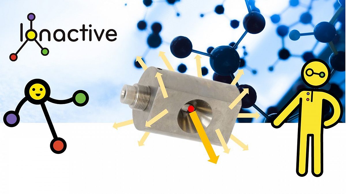
Example of an industrial radiography collimator
For the purposes of this blog we will provide some generic data for this collimator, in reality you would choose a specific collimator for a particular job.
- Designed for Ir-192 and Se-75. We will pick some data for Ir-192.
- Typical radiation conical beam is 60 degrees - this provides a tight collimation down onto the radiographic object.
- The collimator is usually made of tungsten.
- Our example provides 6 HVT for Ir-192.
A full explanation of HVT and TVT can be found in our radiation protection glossary. Suffice to say, that the HVT is the thickness of material that will attenuate gamma (or x-ray) radiation to 1/2 of the pre-shielded value, and TVT is the thickness of material that will attenuate gamma (or x-ray) radiation to 1/10 of the pre-shielded value.
The collimator in question provides 6 HVT. It can be stated that 3.32 HVT = 1TVT (see our resource 'How do I convert TVT (10 value thickness) values to attenuation for Gamma or X-ray sources of radiation?' for a full explanation on this concept).
We note that 6 HVT provides 1.807 TVT. Using the following expression we can calculate the attenuation provided exactly.
\[ Attenuation=10^{-1.807}=0.0156\]
[Ionactive note: we could have converted HVT to attenuation instead by using (0.5)6 = 0.0156]
This attenuation factor can then be applied to the pre-shielded dose rate given above at 1m (41.82 mSv/h). The post-shielded (collimated) dose rate is 0.652 mSv/h (strictly speaking we should note that this is in the horizontal direction towards the barrier being considered).
We can then apply the same inverse square expression as before, but now with this attenuated dose rate (note that dose rates are all in micro Sv/h and distances are in m).
\[d_{2}=\sqrt{\frac{652\times(1)^{2}}{7.5}}\]
This time we see that d2 (distance B in the site radiography graphic ) needs to be > 9.32m to meet the criteria of < 7.5 micro Sv/h. This is a dramatic reduction on the 74.7m calculated earlier and shows the significant advantage of using collimation. A 10 m exclusion zone in all directions, i.e. an area 100 m2, is much more manageable than the previously calculated 5400 m2.
Consideration of the radiographic object intrinsic shielding
Depending on the size, material and orientation of the radiographic object, some credit for its intrinsic shielding may be useful. It is not particularly useful in our example as shown, but imagine if a 'shot' was required in a horizontal direction towards the barrier? In this circumstance little credit can be taken for use of the collimator in the direction of the beam, other than to note the beam will not be 'spread out' along the permitter as it would be if no collimator was used.
We will imagine that the beam is pointing towards the bottom barrier (remind yourself of the setup by referring to the graphic above if required).
We can therefore revert to the pre-collimated dose rate at 1m which was 41.82 micro Sv/h. For the purposes of this example we will imagine that the pipe (radiographic) object has the following properties:
- Steel pipe - nominally 50 cm in diameter.
- Pipe wall is 30mm thick (stainless steel).
Note that the shot is being taken towards the bottom permitter horizontally so the total thickness of steel is 60mm (two walls). If you have a peek at the radionuclide data earlier in this blog you will note that the TVT for Ir-192 and steel is about 43mm. Therefore the intrinsic shielding offered by the pipe is 1.4 TVT (60mm/43mm).
The expression for attenuation is used as follows.
\[ Attenuation=10^{-1.4}=0.040\]
If the attenuation (0.040) is then applied to the dose rate at 1 m (41.82 mSv/h) we see that the attenuated dose rate through both pipe walls is 1.673 mSv/h (1673 micro Sv/h). Following the methodology noted earlier we can then find the distance required to achieve no greater than 7.5 micro Sv/h on the bottom perimeter.
\[d_{2}=\sqrt{\frac{1673\times(1)^{2}}{7.5}}\]
This time we see that d2 (distance B in the site radiography graphic) needs to be > 14.94 m to meet the criteria of < 7.5 micro Sv/h. That is well over 5m more than for our collimator example earlier, even accounting for the approximate 25cm (radius of the pipe) increase in distance provided by moving the collimator to the back of the pipe (and facing the bottom perimeter).
Probably the easiest solution is to move the bottom permitter back 5.5m temporarily for this shot (and do the same the opposite side - for the p permitter - when the time comes). However, imagine there is no room to increase the bottom perimeter distance from the source?
[Ionactive note: Those into industrial radiography might now be shouting loud and clear 'Small Controlled Area Radiography - SCAR'. This is a type of NDT using special projectors, typically rated to 15 Ci (555 GBq) of Ir-192, which are placed directly on the area of the radiographic object which needs testing. This has very tight collimator and, for its size, dramatically reduces leakage / scattered gamma rays in all directions other than the beam direction, allowing for much reduced size of controlled area. Typically these systems have limits on penetration such that Ir-192 at maximum allowable activity is good for about 50mm of steel. A particular advantage is that the source remains within the projector at all times, and is moved remotely to a position where the gamma rays can leave the SCAR projector via a built in tungsten collimator. Since minimisation of controlled area size is the goal, SCAR projectors are usually used with radiation shielding (lead) blankets and other local scatter shields to limit the area further. For the purposes of this particular blog article, we will leave further discussion of SCAR to the expert NDT contractor!].
In our example we will will consider use of some shielding blankets attached to the pipe by bands, in order to reduce radiation dose rate further towards our bottom perimeter. Traditionally lead blankets have been used (and are still used). However, there is more choice available now with composite materials which use tungsten and bismuth elements (some of these are more malleable and easier to handle than lead).
Note that our collimator calculation yielded 652 micro Sv/h at 1m (allowing distance B to be about 9.3m), and our projection through the pipe wall calculation yielded 1673 micro Sv/h at 1m (requiring distance B to be increased to about 14.94m). If we are aiming for something near 9.3m then we are looking to reduce the dose rate considerably such as 652/1673 = 0.389 (this is an attenuation value).
Looking online we note a particular bismuth shielding blanket is reported to provide 1.25 HVT for Ir-192 radiation energy. We can use the following expression to determine the attenuation provided by 1 HVT:
\[ Attenuation(HVT)=10^{(HVT)}\]
So for a HVT of 1.25 we can write:
\[ Attenuation(HVT)=10^{(1.25)}=0.42\]
[Ionactive note: we like to mix things up but don't want readers to be confused, especially if this material is unfamiliar. Carefully inspect the above expression. It might look similar to the TVT expression from earlier, but it is not the same as it is missing a '-' in front of the 1.25. As written this is calculating the attenuation from 1.25 HVT - as required. If a '-' was placed in front of the 1.25 then we would be calculating the attenuation from 1.25 TVT. To check this answer, try the alternative expression for calculating attenuation for a number of HVT = (0.5)1.25 = 0.42, the same answer as expected.]
The use of the single bismuth shielding blanket would yield a dose rate at 1 m of 703 micro Sv/h (rather than the desired 652 micro Sv/h). Using exactly the same inverse square calculations we wrote above in full, it can be seen that the distance B (from source to bottom perimeter, via two 30mm steel walls and the blanket) would need to be about 9.7m (not so far off the 9.3m calculated for the collimator when orientated vertically downwards). Another shielding blanket could be used, but it's just easier to move the barrier out about 0.4m and check with the radiation monitor that this achieves < 7.5 micro Sv/h.
All the considerations so far have been for barriers at ground level (i.e. the source is more or less on the same level as those standing outside the perimeter). Care should be taken to consider what is around the area at height (e.g. perhaps a mezzanine floor overlooking the NDT area). Unless the source is pointed directly upwards (which might require a shielding blanket to be placed on top of the pipe), generally the collimator will do it's job in the same way it does at ground level. As always, if in doubt clear the area (and take radiation dose rate measurements).
Consideration of "flash dose rate" during source deployment & retraction
Here is a a reminder of the open site situation we are analysing, this time we are going to look at dimension A as shown.

Radioactive source in transit between the projector and the radiographic object
The study of this work setup so far reveals that the dimension B is somewhere between 9.3 and 9.7m depending on the orientation of the source, position of the collimator and use of local shielding (such as a bismuth blanket). We have determined that the dose rate at the bottom perimeter is no greater than 7.5 micro Sv/h at these distances.
For the purpose of this blog article we will assume that dimension A is 1.5m less than dimension B (such that the distance from the end of the projector to the source collimator is 1.5m). Therefore, using our smaller dimension for B (9.3m), the dimension A will be about 7.8m.
During the source deployment and retraction the source is push / pulled along the source guide tube and at this point it is neither collimated or shielded in any way. The unshielded dose rate at 1m is 41.82 mSv/h. A simple inverse square calculation will determine the dose rate at the barrier.
\[\frac{D1}{d_{2}^{2}}=\frac{D2}{d_{1}^{^{^{2}}}}\]
In the above expression, D1 is the dose rate (41.82 mSv/h) at 1m (d1) from the source, and D2 is the dose rate at the bottom barrier at 7.8m from the source (d2).
\[D_{2}=\frac{D_{1}\times d_{1}^{2}}{d_{2}^{2}}\]
We can then pop the values in using micro Sv/h for dose rate at m for distance. This gives us
\[D_{2}=\frac{42820\times(1)^{2}}{(7.8)^{2}}\]
So D2, which is the dose rate at the barrier just as the source starts to be deployed (or just prior to entering the projector on retraction) is 687 micro Sv/h ! This is known as an instantaneous dose rate (IDR) and will last (at this magnitude) for no more than a fraction of a second, reducing all the time as the source travels further away from the bottom perimeter and towards the collimator. The magnitude of this IDR dose rate on first inspection seems massive - but some careful consideration is then required.
[For later - we will consider accidental exposures later in this blog. However, even now you may have a clue about what 'might' go wrong. For now it is assumed that the source is moving smoothly and correctly so that the IDR is reducing all the time from first emerging from the projector to the point it reaches the collimator].
If we wanted to be exact (mathematically) there might be some calculus (!) involved since the dose rate is changing rapidly in time and distance. What we do know from experience when monitoring such situations, is that "flash" is a good choice of words, the IDR is over in a flash, so quick that many monitors will, at best, make a noise and you might see a brief higher deflection (analogue monitor) or a higher dose rate (digital). It is more useful to put the monitor into integrating mode (if that function exists) and try and capture the "dose" (not dose rate) during the briefly higher IDR. Most of the time we have seen perhaps a micro Sv captured at most.
Assume the highest dose rate lasts for 0.5 seconds (worse case if the source is moving). At a dose rate of 687 micro Sv/h, that would yield [0.5/3600 * 687] = 0.5 micro Sv (and a bit more from the ever decreasing dose rate as the source moves towards the collimator). This is therefore averaging and we would need to add the contributions from the source fully exposed position (hence would need total exposure time per shot and number of shots per hour etc).
Averaged over a minute we have the following:
- 687 micro Sv/h for 0.5 seconds
- 0.05 micro Sv/h for 59.5 seconds/ Note we are ignoring enhanced dose rate once the source is deployed into the collimator to keep things clearer (hence we are using average background).
The average dose (rate) over a minute can then be seen to be 5.8 micro Sv/h (it will be a little higher due to the contribution of the falling IDR as the source moves towards the collimator). Note - we are just considering the "flash" IDR, not the overall exposure at the boundary from the source once it is located in the collimator.
Overall this is somewhat a nonsense figure and what a radiation monitor can (or cannot) measure is likely to provide a more reliable indication of derived dose rate over a minute or an hour. However, what this analysis does tell us that even significant IDR delivered over short durations can still meet the definition noted earlier i.e. IRR17 ACoP para 298 specifically provides guidance that for industrial radiography, a controlled area will exist where the dose rate exceeds 7.5 micro Sv/h when averaged over a minute (noting this is a minute and not an hour).
What about ALARP? This flash IDR can be lowered in a number of ways. Suggestions include:
- Increasing distance (i.e. increasing distances A and B), meaning moving the barrier further away from the source / projector.
- Moving the projector nearer the radiographic object and using a shorter source guide tube (this effectively increases distance from the source without physically needed to move the perimeter barrier.
- Use the body of the projector, and careful placement of the source guide tube behind it, so that it provides additional local shielding. This would reduce direct beam dose rates but not necessarily radiation scatter.
- Use additional local shielding around the source guide tube nearest the projector (e.g. using bismuth / lead shielding blankets).
Likely occupational doses from site radiography as previously described
Without workload it is impossible to predict occupational doses with certainty. Therefore we will just make up some reasonable data and see what we can derive from it. Note that site radiography is only justified after enclosure radiography has been shown not reasonably practicable. Therefore it is most likely that much of the day to day 'bulk' NDT on production lines etc will be enclosure radiography. Site radiography will never be considered routine and it is more likely to be project based (e.g. a NDT technician may spend two days on site undertaking site radiography for a specific project, and then spend the next couple of weeks undertaking enclosure radiography). We will be looking at occupational exposures from enclosure radiography, based on our featured open-top facility, later in this article.
- Assume 2 shots an hour, 8 hour working day.
- 5 day working week.
- Each shot is 10 minutes.
- The rest of the week is spent setting up shots and other NDT related matters.
- Assume the barrier dose rate is 7.5 micro Sv/h (Ionactive experience - it is likely to be much less than this in most cases).
- Assume the NDT technician stays at the barrier for the entire shot duration.
Occupational exposure for the week from the NDT shots. Total source exposure time is 800 minutes / week (13.34 hours per week). Therefore occupational exposure per week could be 100 micro Sv.
For a 50 week working year this would translate to 5000 micro Sv / year (5mSv /year) which is under the 6 mSv / year whole body dose that would require classified person status (note that all NDT technicians will be classified persons regardless). This is a likely a huge overestimate.
This estimate does not include the "flash" exposure or exposures from carrying the projector to and from the work area. Earlier we estimated each flash exposure to be 0.5 micro Sv, so there would be two per exposure (i.e. 1 micro Sv / shot for flash). Using the data above we see there are up to 80 shots per week, so 4000 shots a year, which could indicate an additional 4000 micro Sv/year. However, monitoring evidence from Ionactive site visits indicate flash exposures per shot are significantly less than our calculation. Remember - calculations are estimates and great for planning but do not replace workplace monitoring and use of dosimetry (including real time active dosimetry). Also note we discussed several methods to mitigate flash exposures and would expect many of them to be used - this alone would lower exposures. Furthermore, our calculation is based on a full workload of site radiography where we are considering a critical person - this being a single NDT technician that undertakes all the shots.
What about non-occupational exposure? This would include, for example, other employees or other persons (e.g. contractors, visitors, possibly members of the public) who are not engaged in NDT operation, but may have some degree of access to the external permitter of the radiography area.
- A project manager visits the NDT technician at the barrier once a week for a 10 minute catch-up. On each visit radiography is taking place. Non-occupational exposure each week would be 2.25 micro Sv.
- A security guard makes 4 tours of the site during an 8 hour working day, and each tour includes 10 minutes within the vicinity of the perimeter. On average it is assumed a radiography shot is taking place during two out of the four visits to area. Weekly non-occupational exposure could be 22.5 micro Sv over a five day week where radiography takes place. [Note. Whilst this might be acceptable for a few weeks of NDT, more than 13 weeks of this work would move this person over the 300 micro Sv/year whole body dose constraint for those not involved in work with ionising radiation].
There are many more examples we could look at. The important thing to note is that ALARP must be applied and regular access to the permitter by those not working with ionising radiation would suggest lowering the dose rate below 7.5 micro Sv/h.
We will now consider occupational exposures from enclosure radiography.
Occupational exposures from enclosure industrial radiography
[Ionactive comment. A modern NDT gamma source enclosure with shielded roof and HASS source security provision (i.e. projector does not need to be taken to and from the enclosure each day), should not need to yield occupational exposures above background. They can be engineered such that exterior dose rates are < 1 micro Sv/h which tends towards background (nominally 0.05 - 0.1 micro Sv/h) at 10 cm from the surface. For this blog we will consider our open topped NDT enclosure which might not be ideal in all cases, but was reasonably practical for the purpose it was designed for at the time].
A reminder of the layout of the enclosure.

Open topped NDT radiography enclosure (side view)

Open topped NDT radiography enclosure (top view)
Enclosure shielding materials
Where the enclosure is physically small and / or where space is restricted, it is likely that lead will be the preferred shielding material.
For this blog we will assume that the TVT for lead with Ir-192 is 20mm (note: this does vary and some references report as low as 10mm).
[Ionactive comment: For those that want to delve in deeper and consider TVT and its discrepancies, you may want to read our resource article here: 'How reliable is TVT (10th Value Thickness) in radiation shielding calculations?'. The problem with references is that they propagate around the internet and often don't show where they are derived from - this is why we resist putting references into these blog articles unless we are certain they are reasonable. In the case of Ir-192 with lead, the TVT is reported as 20mm in table 6.4, page 6-15 (Exposure & shielding from external radiation) from the publication - 'Handbook of Health Physics and Radiological Health' (3rd edition). Our link noted above goes into TVT in some detail, but basically considers matters such as buildup, the '1st TVT' (TVT1) and the 'Equilibrium TVT' (TVTe). In the publication 'Design and Shielding of Radiotherapy Treatment Facilities' IPEM Report 75, 2nd Edition TVT1 is reported as 11mm and TVTe is reported as 19mm (table 10.4, page 10-11). It is no wonder that references (or those quoting them) may pick and choose. What is important is real world data, and NDT technicians will know very well what actually works! The same discussion can be made for other materials such as concrete - but we will park this matter for another day].
We know that 3 TVT (60mm lead for Ir-192) will provide a 1/1000 fold reduction, so considering the dose rate at 1 m from the projector (see earlier), this would reduce 41.82 mSv per hour (at 1m) to 41.62 micro Sv/h. If the average wall distance was at least 3 m from the source, and the source was in the middle of the enclosure (and its minimum distance from the wall restricted by engineered means), this dose rate could be reduced further by 1/32 (1/9) which would give us about 4.62 micro Sv/h. Then introduce the collimator from the earlier discussion on site radiography (6 HVT = 1.807 TVT = attenuation of 0.0156), and the dose rate on the external perimeter of the enclosure drops to 0.07 micro Sv/h (i.e. background). So Lead is possible, but consider the following:
- To be cost effective (and structurally sound) lead is only really suitable for smaller enclosures.
- The dose rates calculated above were for 370 GBq (10 Ci) of Ir-192, so the enclosure would need a site based license (or equivalent) for maximum allowable activity.
- Restricting source distance from wall needs engineered means (e.g. short source guide tube), but this might restrict the size and orientation of the radiographic object.
- Use of the collimator will rely on human factors (i.e. using it) - this needs to be carefully considered and justified, or the work may not meet enclosure radiography conditions (might still be considered site radiography).
- Access to a relatively small enclosure would usually require a sliding door rather than a maze. Suppose the lead in the door was 1.8m (high), 2.5m (width) to allow movement of radiographic objects in and out, and 0.06m (60mm) thick then the door (without its steel frame) would have a volume of around 0.27 m3. Since lead has a density of 11400 Kg/m3, the minimum mass of our door would be around 3000 kg (3 tonnes). That is a lot of door to slide back and forth, even with the help of pneumatic systems.
So lead is possible - but not for our open top enclosure. So let's now look in more detail at its shielding.
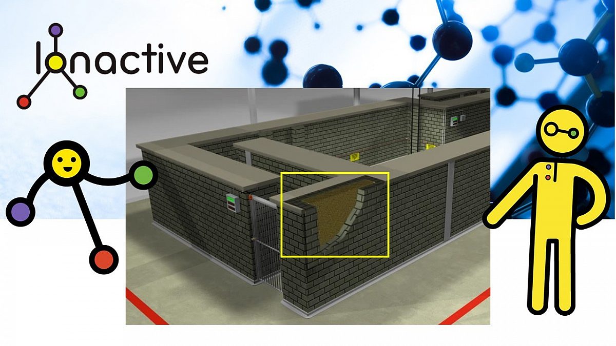
Open top enclosure industrial radiography - shielding
The shielding for this facility was designed with the aid of computer modelling software. For this blog we will keep things simple and consider just empirical factors. The shielding detail are as follows:
- Inner wall 21.5 cm of 1.9 density concrete block.
- Middle area 46 cm of 1.7 density compacted sand.
- Outer wall 21.5 cm of 1.9 density concrete block.
The height of the walls is 2.5 m (we will discuss potential sky shine later in this blog).
Standard density concrete is generally taken to be 2.35 g cm-3 (we call this 2.35 density, where water would therefore have a density of 1).
Let the TVT for Ir-192 at 2.35 density concrete be 14.7 cm. This is taken from table 6.4, page 6-15 (Exposure & shielding from external radiation) from the publication - 'Handbook of Health Physics and Radiological Health' (3rd edition).
To a good approximation we can correct for density in concrete by considering ratio of densities. Therefore the TVT for 1.9 concrete with Ir-192 is inferred to be - 18.2 cm. For simplicity, we will assume the sand is equivalent to concrete at 1.7 density (whereas the actual shielding model considered sand explicitly). So we can infer that 1.7 density sand provides a TVT of 20.3cm.
[Ionactive comment. Use density ratios with care! Generally at reasonably high photon energy (> 511 keV) you can ratio densities to adjust TVT of the same material type. Don't ever try this using say lead and concrete density ratios as it will not work. For a fuller explanation please have a read of the following Ionactive resource: Can I use density ratios when working out radiation shielding thickness? Since we are increasing TVT with lower density, at worse we will overestimate the shielding required].
With the above data accepted, we can determine the total TVT provided as follows:
Inner wall - TVT is 21.5/18.2 = 1.2 TVT
Middle (sand area) - Total TVT is 46 / 20.3 = 2.3 TVT
Outer wall - is 21.5/18.2 = 1.2 TVT
So the total TVT offered by the wall for Ir-192 is 4.7 TVT.
Using the attenuation equations earlier, we can see that 4.7 TVT has an attenuation factor of 2 x 10⁻⁵ (0.00002) (Note: attenuation in this example = 10-4.7). We will now examine this attenuation in our shielding example.
[Sanity check on TVT - attenuation. Suppose we have a dose rate of 1,000,000 micro Sv/h. An attenuation of 0.00002 (2 x 10⁻⁵) would reduce this to : 20 micro Sv/h].
We will now position a 370 GBq (10 Ci) Ir-192 source completely unshielded inside the enclosure and consider the shielding performance in terms of dose rate outside the wall (sky shine over the top will be considered later). The next graphic provides some dimensions we can work with.

Calculation position with dimensions
Occupational exposure dose rates through enclosure shielding
We note that the distance between source and calculation point is 3.9m and this is through 4.7 TVT (0.00002 attenuation as shown above). We have specified the dose rate for 370 GBq (10 Ci) of Ir-192 as 41.82 mSv/h at 1m earlier in this article. Following the simple calculation methodology shown earlier we apply both the inverse square law and attenuation.
\[ Wall dose rate=\frac{1}{3.9^{2}}\times 0.00002\times 41820=0.055\mu Sv/h\]
Therefore dose rate is reduced to background levels through the wall. Note that the dose rate without the shielding wall would be 2750 micro Sv/h at 3.9m from the source. Also note that the wall shielded dose rate is for an unshielded and uncollimated source with no radiographic object.
[Ionactive comment: Depending on workplace circumstances, this dose rate could also be used to calculate non-occupational exposures to those not involved in the radiography].
Occupational exposure dose rates - shy shine (scatter)
We now consider dose rates outside the enclosure which are from sky shine (scatter) over the top of the shielding wall. The values reported below are from actual measurements taken around the enclosure. We will compare some of this data with calculated values - however we will not detail the calculation methodology for sky shine as it is way more complicated than anything so far considered and outside the scope of this article. For a more in depth consideration, please see this Ionactive resource: Skyshine – radiation scattering around and over shielding (we will use a picture from this resource which is shown below).

Photon radiation skyshine over the top of a shield
The following graphic illustrates some key dimensions for consideration outside the enclosure (these are not to scale - just believe them!). They show:
- 3m from dose point (green circle) to red perimeter line (no work above 2.5m in this zone).
- 3m from the red perimeter to the red platform (critical worker is located such that the trunk of their body is 3m above the ground).

Enclosure external dimensions - for dose rate illustration
The data shown below is real dose rate measurement results from skyshine for a 370 GBq (10 Ci) Ir-192 source. In this blog article we will only consider dose rates in one direction (as shown above). We could limit dose rates in other directions with a collimator, and this would reduce some of the radiation over the top of the bay. However, ultimately this facility is for radiography of large bore pipes with a panoramic collimator. This type of collimator will:
- Reduce radiation exposures either end of the bay.
- Reduce the scatter above the enclosure either side of our (green) measurement spot.
It follows that if we are aligned with the source (with or without panoramic collimation), the dose rates being considered will tend to a maximum.

Dose rates outside the enclosure - fully exposed source
Some initial observations.
- With the exception of the 3m height measurements, peak dose rate is somewhere between 4-6m from the shielding wall (there is a shadowing effect close to the shielding wall).
- For the 3m height measurements, highest dose rates are near the wall and drop of with distance (not surprising as the wall offers less of a shadowing effect at this height).
- Notwithstanding exposure time (considered shortly below), dose rates are significantly above 7.5 micro Sv/h in areas where employees external to the enclosure may work - this is not acceptable (or ALARP).
- Always consider skyshine and monitor further enough away from the shielding wall to ensure you capture the highest potential dose rates.
- The observations above could also be used for assessment of non-occupational (i.e. for other persons not involved with work involving ionising radiation).
We now consider the scenario where the Ir-192 source is inside an 18mm steel wall wellhead pipe. For this facility 18mm steel is the smallest thickness available, more common wall thicknesses available vary between 25-50mm.

Dose rates outside the enclosure - with 18mm wall thickness wellhead
The dose rate measurement results show an expected reduction - the overall pattern of dose rates is similar to the completely unshielded source. We now see that waist and head height dose rates are substantially below 7.5 micro Sv/h. The working at height (3m) dose rates, from 6m onwards are substantially < 7.5 micro Sv/h, and whilst there is no intention of working at height close to the enclosure (i.e. inside the red permitter line), dose rates 4-5m away are still significantly above 7.5 micro Sv/h. This would be likely rectified with thicker walled wellheads.
For example, suppose the steel wall thickness was 32mm (i.e. 32mm-18mm = 14mm thicker). We note from earlier that the HVT for Ir-192 with steel is about 13mm. Therefore, we would expect, at the very least, for a 1 HVT reduction in dose rates (i.e. at least 50% reduction in the dose rate values noted in the table above).
However, it was decided to build a shielding canopy to be manoeuvred over the radiographic object. This was built to run on rails either side of the wellhead to ensure that its use became an integral part of the radiographic procedure, rather than an afterthought. Early on it was decided that the canopy would be used for wellheads with wall thickness below 35mm steel, however once work in the enclosure was underway a decision was made to use the canopy regardless of wellhead wall thickness (this reduces the risk of the canopy not being used when required).
Have a look at the dose rate data below with the shielding canopy in place over the 18mm wellhead. The construction details of the canopy is given afterwards.

Dose rates outside the enclosure - with 18mm wellhead and shielding canopy
Let's explore the shielding canopy in more detail.
Much earlier in this article we have specified TVT Steel as 43 mm for Ir-192. We have also specified TVT lead as 20 mm for Ir-192.
The shielding canopy is made from 12mm steel, 6mm lead another 12 mm steel. So we have 24mm steel in total and 6mm lead. The attenuation provide by this canopy (in theory) can be expressed as follows below. [Note: In practice the shielding canopy should do better (or at least as well as) the calculated attenuation since it is dealing with lower energy scatter and not just primary radiation from the source].
The attention expressions for the lead and total steel present can be expressed as follows:
\[ Attenuation(Pb)=10^{-\left(\frac{6}{20}\right)}=0.501\]
\[ Attenuation(Fe)=10^{-\left(\frac{24}{43}\right)}=0.277\]
The total attenuation (lead plus steel) is the product of the values which is 0.14 (rounded).
How does this compare with the measured values when using the canopy? Pretty close it seems! Try using the 0.14 attenuation and applying to the pre-canopy table above.
[Ionactive comment: Throwing some additional shielding over the top of a radiographic object would be more appropriate for site radiography than enclosure radiography. However, in our example the canopy is engineered for a specific type of work and runs on rails either side of the laydown markers for the wellhead. Just as 'search and lockup' (looking for a person left in the enclosure upon exit) is a procedure (with associated human factors), so is the use of the canopy. Therefore in our specific example the canopy is an integral part of the process and use of the facility and so enclosure radiography is justified. This conclusion will be investigated further below when we consider potential exposures to those outside the enclosure when radiography is underway].
Likely exposures to those inside the enclosure setting up the radiographic shot and operating the system from the control panel outside the enclosure
This is clearly occupational exposure.
This can be estimated by looking back at our data for the radiographic projector and our discussion on open site radiography. However, the actual exposures will be much less due to:
- No exposure to flash dose rate during source deployment and retraction.
- Exposure very near background around the permitter of the enclosure and ground level (including the control desk area) during the exposure.
Our estimate (based on a workload which will be described below) is that a radiography technician operating only this facility for 8 hours / day, 5 days a week for a full working year, would not receive an occupational exposure measurable above background. This is an interesting conclusion when assessing exposures to other persons some distance from the enclosure which we will now consider.
Likely exposures to those working away from the enclosure and who have nothing to do with the radiographic work taking place in the enclosure
We will first consider some workload data. In the last table of data we have shown that with the shielding canopy, regardless of type of wellhead, instantaneous dose rates (IDR) are significantly < 7.5 micro Sv/h. With the 18 mm (wall thickness) wellhead, the highest IDR (in our direction of interest) is 3.5 micro Sv/h (at 3 m height in the red perimeter restricted zone). The highest waist level dose rate is 0.4 micro Sv/h (at 4 m from the enclosure wall) and the highest permitted working at height (3 m) dose rate is 0.6 micro Sv/h (at 6 m from the enclosure wall). This data might be easier to digest by simply looking at the last table of dose rate data provided. We will use these values to explore exposures in these areas.
Workload
It was determined that a panoramic shot for the 18 mm wellhead would need 100 Ci.min. Therefore, the duration of each shot will be 10 mins (based on the 370 GBq / 10nCi Ir-192 source).
It was further determined that each wellhead "weld" would need 3 shots, so total duration of radiography was 30 mins per wellhead.
Radiography technicians determined that the total set up time (i.e. craning wellhead into the enclosure, setting up the radiography, performing the radiography and craning wellhead out of the enclosure) would take 2 hours (120 minutes). Therefore the radiography 'source exposure' period is 30 minutes in every 120 minutes.
At full working rate there could be up to 90 minutes of radiography source exposure time in an 8 hour working day (i.e. 3 wellhead over 6 hours plus 2 hours of breaks / rest / maintenance / paperwork etc).
[Note: A typical 50mm walled wellhead will need up to 250 Ci.min, which would mean an exposure time of 25 minutes per shot. However, the additional attenuation of scatter from this well head will be significant. The additional steel is 32mm, so the additional attenuation is 10(-32/43) = 0.18 (revisit earlier if you need to consider how this works). Whilst the exposure time for the thickest wellhead is 2.5 times more than for the thinnest wellhead, this is compensated by the additional attenuation (nearly 5 times more). For this blog article we will just stick with the thinnest wellhead considerations].
Dose assessment
We will present the dose assessment as a weekly exposure - this can be multiplied by 50 to provide an annual value.
Total exposure time in a week is [5 days / week x 90 minutes / day exposure] = 450 minutes / week (i.e. 7.5 hours / week).
- Consider Person CP1 standing 4 m from the wall (see picture below). If they were located at this point for the whole week the weekly waist (trunk of body) exposure would be [0.4 micro Sv/h x 7.5 hours / week] = 3 micro Sv/week (150 micro Sv/ year). Exposure to the head (eyes) would be [0.5 micro Sv/h x 7.5 hours / week] = 3.75 micro Sv/week (188 micro Sv/ year). This assumes 100% occupancy which is not realistic for this work location (if an admin office was located at this position it might be more valid). So actual exposure will be substantially less than this.

Dose assessment for critical person (CP1)
- Consider Person CP2 standing 6m from the wall where their trunk position is at an average 3m above the floor (see pictures earlier). If they were located at this point for the whole week the weekly waist height (trunk of body) exposure would be [0.6 micro Sv/h x 7.5 hours / week] = 4.5 micro Sv/week (225 micro Sv/ year). Since the height may vary, and due to lack of further data, we will attribute eye dose to the same value (225 micro Sv/year). The trunk dose is 75% of our dose constraint of 300 micro Sv/year (whole body dose). However, this assumes 100% occupancy of the critical person in this position which is not realistic. Actual weekly / annual exposure is likely to be considerably less than predicted here.
This blog article provides examples of an assessment process - if this was for real similar assessments would take place all around the enclosure. Before we move on it is appropriate to briefly consider again the occupational exposure potential of the radiographic technicians who are working outside the enclosure, in the region of the control panel.
Consider the picture below and the analysis which follows.

Dose rates in region of control panel - 18mm wellhead and canopy
- The radiography technician is standing / sitting 0.5m from the wall next to the control panel. The average dose rate in the area was measured as 0.15 micro Sv/h (close to background). If they were located at this point for the whole week (after setting up each radiographic shot) the weekly whole body exposure would be [0.15 micro Sv/h x 7.5 hours / week] = 1.13 micro Sv/week (57 micro Sv/ year). This assumes 100% occupancy at this point which is a more reasonable assumption compared to those who are not occupational exposed (since they should be in close control of the work at all times).
The IDR specified above was 0.15 micro Sv/h - this is so close to background that many radiation monitor users would quote background (especially if the monitor had an analogue scale). Therefore the quoted exposure over a year is unlikely to be distinguishable from background.
What is interesting about this result is that likely exposure to the radiography technician outside the enclosure is less than the calculated exposures to those - not occupationally exposed - further away from the enclosure. Skyshine - it has a lot to answer for! That said, we have shown that for compliant use of the enclosure, all exposures outside the enclosure are less than our 300 micro Sv/year whole body dose constraint.
[Ionactive comment: a word about radiation monitoring. In this blog article we have used instantaneous dose rate (IDR) measurements to assess likely annual exposures for a number of scenarios. However, whilst the article is based (more or less) on a real facility, the monitoring conducted was significantly more rigorous than might otherwise be suggested. For example, accumulated dose measurements were used to support IDR measurements. A radiation monitor was set to measure 'dose' and not 'dose rate'. Over a complete morning of representative radiography the monitor accumulated a dose value - this was background corrected with another monitor placed in another building. This accumulated value was then integrated over a week and / or year. Furthermore, during the enclosure trials and after commissioning, passive environmental dosimeters were placed around the enclosure including positions at 3 m (as illustrated earlier for CP2). Annual predicted exposures from accumulated dose were very similar to those calculated from IDR measurements. This is not surprising since the exposure periods were long (10 - 25 minutes) so there was little variation of dose rate over time which an IDR based measurement might miss].
What about exposures at the entrance gates to the enclosure? Note the following picture and the analysis which follows.

Dose rates in region of the interlocked gates - 18mm wellhead and canopy
If you look at the above figures and picture you can see that the annual 'gate' whole body dose is predicted to be 57 micro Sv/ year. We have already stated that this is unlikely to be measurable above background. This would also assume 100% occupancy which for the two gate locations is nonsense. For 10% occupancy there is no accumulated exposure measurable above background in these areas.
We have arrived at the end of this section considering occupational exposures (and also non occupational exposures to other workers). We are now ready to consider accidental radiation exposures in industrial radiography - considering both open site and enclosure examples.
Accidental radiation exposures
The headline title of this blog article is not meant to be misleading! However, we should state clearly that we are going to consider:
- Radiation accidents.
- Unintended or unplanned exposures (which could be a subset of radiation accidents).
Thankfully unintended / unplanned exposures which do not lead to radiation accidents are more common than actual radiation accidents. Before we get stuck into this section it is important to define what is meant by radiation accident (UK perspective).
Regulation (2)(1) of IRR17 defines radiation accident as follows: '...an accident where immediate action would be required to prevent or reduce the exposure to ionising radiation of employees or any other persons...'.
A radioactive source which fails to retract on demand (during site or enclosure radiography) is a radiation incident (to be mitigated), but not necessarily a radiation accident (but could become one if the mitigation is poorly implemented). In the above mentioned enclosure - if a power cut stopped source retraction from completing, the source could be made safe with negligible employee exposure by using the manual motor winding handle (located outside the enclosure). This is a radiation incident, but it is not a radiation accident.
We can dig deeper to consider what would be a radiation accident (or how to avoid the accident). Paragraph 121 of the IRR17 ACoP (Regulation 9(2)) states '...Where there is a risk of receiving an exposure within a few minutes that could cause deterministic effects, it should be reasonably practicable for employers to install engineered search and lock-up systems into the exposure control system...'. We have described such a system installed into the enclosure bay considered earlier - this is absolutely designed to avoid radiation accidents.
Following the ALARP principle indicates that dose mitigation is worthwhile at most points on the likely exposure scale (unless you can demonstrate that ALARP has been achieved and nothing more can be reasonably done). However, the above wording implies that a sensible use of the definition of a radiation accident is where immediate actions are required to (in the following order):
- Absolutely avoid deterministic radiation effects.
- Mitigate radiation exposures to below legal limits (effective and equivalent doses).
- Mitigate radiation exposures lower still, to ALARP levels compatible with the nature of the radiation accident (i.e. significantly below legal dose limits).
The use of 'radiation accident' / 'incident' will be used interchangeably to describe the scenarios which follow. We will leave you to decide which events are truly radiation accidents as defined in IRR17.
Route causes - why radiation accidents in industrial radiography?
There are some useful publications which have analysed this question. One such publication is IAEA Safety Report Series 7 - Lessons Learned from Accidents in Industrial Radiography (opens in a new window). This publication is from 1998 and we would hope that significant improvements in NDT safety have been made since. Accidents of the type described shortly are thankfully very rare in the UK. However, we have seen poor practice whilst on work trips to the Gulf well into the 2010's, so across the world there are still significant improvements to be made.
Another resource to take a look at is here: IAEA - The information channel on nuclear and radiological events (opens in a new window). At the time of writing this blog we note that the latest event was posted 6 December 2023 'Worker Exceeded Annual Whole Body Dose Limit' and involves a 2.33 TBq (63 Ci) Ir-192 source. This incident was due to poor training and supervision and resulted in a whole body dose of 7.5 mSv whole body dose and 258 mSv to the extremities.
[Ionactive note: We really do encourage you to take a look at the IAEA events database from time to time. Whilst it was designed originally for recording nuclear plant incidents (and such events are still recorded), you will note that many of the events are non-nuclear and are mostly concerned with accidental exposures during industrial radiography and use of ionising radiation in medical applications. This is where significant radiation exposures are being received, not from the nuclear industry].
Generally industrial radiography radiation accidents appear to have the following route causes (these are written in descending order of likely occurrence).
- Poor regulatory control.
- Not following procedures.
- Poor (or no) training.
- Poor (or no) maintenance.
- Poor (or no) use of monitors & dosimetry (this features in most of the other categories and examples below).
- Human error.
- Faulty radiographic equipment.
- Equipment design issues.
- Deliberate poor practice.
We will explore these in a little more detail and supply a working example of each. After this we will return to our own previously described sources and look at some accident scenarios involving them.
Poor regulatory control
Thankfully poor regulatory control is something the UK does not need to worry about (much). Radiation safety in the UK is highly regulated - probably gaining a step change in standards with the Ionising Radiations Regulations 1985 (IRR85), which were driven in part by ICRP 26 and relevant EU basic safety standards directives. The most up to date UK regulation is based on the Ionising Radiations Regulations 2017 (IRR17) together with other supporting legislation dealing with protection of the environmental (use of radioactive materials and creation of radioactive waste) and the transport of radioactive materials. Ionactive has produced guidance which might be useful to our readers (both will open in a new window).
- Ionactive guide to the Ionising Radiations Regulations 2017 (IRR17)
- Ionactive guide to the UK Environmental Permitting Regulations (radioactive material and waste)
Ionactive has experience working in places around the world where there is little (or no) obvious radiation protection regulation. In some cases at best there might be a single 'radiation safety' person within a government department. Therefore reliance is often placed on the radiography contractor who might be based locally, or alternatively from another country. There is often no regulation or oversight of their equipment, radioactive or x-ray source , training, contingency arrangements etc. If the contractor is exemplary in every way, then excellent - this is usually the case where a big multinational company (e.g. oil and gas) brings in their own approved contractor to undertake NDT. However the evidence shows that without regulation poor practice propagates as indicated in the following examples.
- In the early 80's a company lacking regulation from the country where it resided was undertaking NDT work with Ir-192. The operator of the radiography system had no training and no concept of radiation safety. The cable drive of the source projector failed leading to the source not being retracted into the shielding. The operator reported this to their supervisor, but since the failure did not appear to impact adversely on the ability to conduct NDT, they were told to continue (!). He soon suffered from deterministic radiation effects (radiation sickness). Later evaluation of his dosimeter showed he had likely received 5 Sv (5 Gy) over a period of two weeks.
- In the middle 70's a 7400 GBq (200Ci) Ir-192 source was being used to radiograph welds on a pipe laying barge. An assessment of a contactor film badge revealed a dose of 30 mSv and evidence of Ir-192 contamination. During the investigation it was found that a pipe being tested had rolled and crushed the lead collimator on the end of the source guide tube. The compression on the tube meant that the source could not be retracted back into the shielded projector. The contractor used a welding torch (!) to melt the lead collimator to free the source. The source, now damaged, was retracted and it was used continuously until the contamination on the dosimeter was reported.
In both examples the radiography contractors had no license or permit, no regulation, no contingency arrangements and no processes to follow. There was no regulator to answer to and it appears they made things up on the spot when faced with a problem.
Not following procedures
Despite the fact that many radiography contractors around the world are regulated, there is still evidence that some do not follow the procedures they have written (or procedures written for them). Here are a couple of examples from this category.
- During 1993 a radiographer received 3 Sv (3 Gy) to his right hand and 12 mSv whole body dose following an incident. He was working with a 3600 GBq (97 Ci) source of Ir-192. Something was clearly wrong as his real time dosimeter (EPD) was alarming - however, for some reason he did not respond to this (as required by the procedures he was supposed to follow). He also appears to have ignored an off scale indication on his radiation survey monitor. He tried to correct the problem rather than contact the company radiation protection officer (also a requirement of the company procedures he failed to follow).
- During 1994 an industrial radiography technician was working on a night shift with a 780 GBq (21 Ci) Ir-192 source. He appears to have had difficulty in locking the projector into a safe state. His alarming EPD (active dosimeter) was reading off scale, but a defective radiation monitor was not responding. To achieve the projector locking he desired, he hit the device with a hammer. He then (presuming the projector was now safe) left the area to obtain a working radiation monitor. Upon returning to the radiographic area he noted the alarming EPD was still off scale, the replacement radiation monitor was also not working, and the locking mechanism on the projector was still defective. His passive radiation dosimeter indicated 8.5 mSv whole body exposure.
Clearly procedures were not followed in the above examples. However, they appear to demonstrate lack of training and maintenance of equipment.
Poor or inadequate training
Many of the examples in this section are interrelated - so poor or inadequate training will be a result of other failings (such as lack of regulation, management or supervision). The following two examples are particularly shocking.
- Mid 1980's an employee was exposed to high doses of ionising radiation from an Ir-192 source. They were not qualified as a radiography technician, they were unskilled local labour who had been contracted to do this work (which involved radiography of pipe welds on a gas pipeline). The labourer had received no training in radiation protection, and did not understand / appreciate the purpose of personal or area radiation monitoring. Unbeknown to labourer, following the first exposure the radioactive source failed to return safely to the shielded projector, instead they continued with the work regardless. On completion of the work it was discovered that some of the radiographic films were "black", indicating
- high radiation exposures. Upon investigation it was established the individual had received 24 Sv (24 Gy) to the hand and 2-3 Sv (2-3 Gy) to the whole body.
- During a radiography procedure a technician did not realise that a radioactive source had moved from a safe position during the movement of a source projector. The technician was wearing an active dosimeter which went into alarm, but they decided the dosimeter was defective so turned it off for the remaining duration of the work (!). Later investigations showed that the technician had received 30 mSv whole body dose.
Poor or inadequate maintenance
Thankfully the latest radiographic source projectors provide fail safe features to minimise unplanned exposures by ensuring the source remains securely attached to source drive cables etc. Whilst all equipment should be maintained as per manufacturer guidance, poor maintenance of older equipment can be particularly troublesome.
- During radiography the source assembly of a projector become stuck in a cut in its guide tube. This damage had not been detected prior to use. The radiographer disconnected the tube and shook it vigorously with both hands in an attempt to release the source. Three fingers on left hand and two on the right hand were exposed up to 8.8 Sv (8.8 Gy), requiring the injured fingers to be amputated. Estimated whole body dose was 0.1 Sv (100 mSv).
- A radiographer and assistant were working with a 3000 GBq (80 Ci) Ir-192 source. Upon completion of radiography the assistant dissembled the equipment, placed it in a truck and drove back to base. Upon removing projector from truck and carrying to storage facility, the source assembly fell out of the projector and onto the floor, as the unit was tilted and placed onto a shelf. A radiation alarm in the storage area immediately sounded and action was taken to recover the source (safely it appears). The subsequent investigation revealed that the unit had undergone no recent maintenance, and the spring loaded latch designed to secure the source in the shielded position was defective (the latch was jammed in the unlocked position). In addition, the projector shutter position was 'on' and the dust cap was not installed. A combination of poor maintenance, defective equipment and poor training resulted in the radioactive source falling to the floor. In this particular case use of the radiation alarm probably saved the individuals from further exposure as they reacted quickly and correctly to the situation upon the alarm sounding.
Human error
Human error is often near the route cause of many accidents, even where the apparent cause may on first inspection be a mechanical defect or a design fault. It is usually human factors which decide where maintenance takes place, and how safety is designed into work equipment. Despite rigorous training, fatigue or distractions can lead to human errors (which is why the engineered safety features of enclosure radiography is designed to reduce human factor based accidents).
- A contractor was undertaking radiography using a 3000 GBq (80 Ci) radioactive source. The NDT was being performed on a number of heat exchanger welds. During this process the source guide cable came into contact with a welders unshielded HV cable. The HV grounded through the guide tube and source projector - the heat generated melted the guide tube such that the source could not be returned to the safe shielded position. The source retrieval was successful but still resulted in 1.5 mSv whole body dose to the radiographers involved. Of note, the report indicates that one of the radiographers received thermal burns to their hands as a result of the high current carried in the conducting guide tube. Arguably this is as serious as any ionising radiation exposure and HV can be fatal. The report indicates that the radiographers had earlier noted the defective welding cables but nothing was done to make them safe.
- Upon completion of radiography, a radiographer placed a source projector on the rear bumper of his work vehicle. He then drove back to the office forgetting he had not safely packed the projector. He only noticed the equipment was missing after arriving back at the office. He retraced a route back to the worksite with a radiation monitor on the dashboard. He located the equipment on the roadside, the source assembly had become dislodged from the shielding during the fall onto the road.
Faulty radiographic equipment
- A radiographer was using a 2700 GBq (73 Ci) Ir-192 which failed to retract into the expected safe shielded position. This was caused by a 'crank-out' failure where the gearbox of the winder separated from its inner liner. The situation was recovered by the operator cutting the housing near the gear box and then pulling the source (via the inner drive cable) manually back into the desired shielded position. No overexposure occurred and this incident was referred to the regulatory authority.
- Whilst a source projector was being wheeled on a trolley back towards it's storage room, a 200 GBq (6 Ci) Ir-192 source fell out due to a defective locking mechanism (the radiographer did not notice this). Two canteen workers found the source and picked it up - seeing the trefoil symbol they reported their discovery. Dose to their fingers was estimated at 8 Gy, whole body dose 200 mSv.
Equipment design issues
Thankfully this is a rare occurrence in equipment designed over the last few decades, and even earlier still.
- During 1977 a 260 GBq (7 Ci) Ir-192 source fell from a pneumatically operated exposure device - the radiographer did not realise this due to a faulty radiation monitor. A construction supervisor picked up the source, thinking it was a component of a nearby crane and placed in left pocket. After trip home on bus with six work colleagues he become nauseous and vomited. He removed his shirt which was then located near his wife and son. An investigation the next day eventually lead to the active source being removed from the bedside table of sick supervisor. 10 Sv (10 Gy) received to right hand (amputated). Up to 100 Sv (100 Gy) received to chest wall (needed skin grafts). Child received 100mSv and wife 170 mSv (whole body dose). Regulatory authority banned the use of this particular device (due to design flaws) but also noted failure to use a working monitor contributed to the large exposures received by the supervisor and family (and also members of the public on the bus).
- During 1989 a 1400 GBq (38 Ci) Ir-192 source assembly disconnected from the source drive cable, within the guide tube. The radiographer successfully recovered the source into a safe shielded location (but still received 2.2 mSv whole body dose in the process). After investigation it was discovered that the hook mechanism which connected the source assembly to the drive cable could be easily disconnected. This design flaw is not common since it is recognised absolutely that the reliability of the connection between source and drive cable is of vital importance.
Deliberate poor practice / wilful negligence
It appears to be rare (thankfully) that a worker would deliberately be negligent with a radioactive source, especially if their training or knowledge highlights the significant harm from doing so. Unfortunately the same cannot be said for a small group of employers who would readily allow such bad practice due to work pressure, lack of resources, time constraints, contractor fees etc. Furthermore, there will be others with no particular knowledge of the risks who will steal equipment containing radioactive sources with little awareness of the health effects to themselves or others they pass the equipment on to.
- In the early 1990's an organization deliberately allowed untrained individuals to perform industrial radiography at their site (this was a breach of known procedures and local rules). Following a radiographic disconnection between source assembly and drive cable, the untrained individuals did not act as would have been expected if trained and following procedures. This resulted in a whole body exposure of 20 mSv to each, with the hand of one individual having a calculated exposure of 900 mSv. No individual was wearing an alarming dosimeter and they had received no training on how to use a radiation dose rate monitor.
- During 1991 a 26 GBq (0.7 Ci) Ir-192 source assembly stored in a lead shield was stolen from a storage pit, together with the unloaded source projector. The theft was unnoticed for several days and the authorities did not identify the thieves. The source assembly changed hands several times and ultimately was discovered at a scrap dealers shop. Scrap dealers estimated whole body dose was 200 mSv, members of the public were estimated at a maximum of 35 mSv.
This ends our examples of radiation incidents / accidents involving industrial radiography. Whilst we have only provided a few it seems apparent that:
- Most of the incidents involve open site radiography, rather than enclosure radiography.
- Most incidents involve use of radioactive sources rather than radiation generators (e.g. x-ray sets).
- By far the most popular radionuclide involved is Ir-192.
- Compared to our own industrial radiography case studies given earlier, the activities are much higher. Our own Ir-192 source under consideration is 370 GBq (10 Ci), whereas Ir-192 sources feature in the above examples were up to 7400 GBq (200 Ci).
We will now complete this blog article by returning to our own featured radiography source projector (for open site radiography) and radiography and consider the dose rates / doses involved in certain accident / incident scenarios.
Accidental radiation exposure / incident - site radiography
Firstly, a quick reminder of what we are working with.
- 370 GBq (10 Ci) Ir-192 radioactive source in a projector.
- 41.82 mSv/h at 1 m from totally unshielded source.
Note that the activity here is considerably less than some of the sources featured in our review above. Recall that you can scale the activity and dose rate - so a 3000 GBq (80 Ci) Ir-192 will yield a dose rate of about 335 mSv/h at 1m (all other things being equal).
A reminder of the workplace situation is given in the picture below.

Representation of open site industrial radiography
We are not going to feature source recovery methods in any significant detail (qualified NDT technicians will know all about this). We will consider the concepts involved, and in particular when they are not followed. Generally where a high activity sealed source (HASS) is in an uncontrolled or unsafe situation the following concepts will always be employed:
- Minimise exposure time so far as is reasonably practicable.
- Maximise distance from the source so far as is reasonably practicable.
- Use shielding (either local shielding placed over the source, or shielding features such as staying behind the source projector if this provides a shielding advantage).
Even for very high activity sources (i.e. much higher than the 370 GBq / 10 Ci source featured in our example), the literature review shows that whole body exposures leading to deterministic radiation effects (radiation sickness) are still thankfully rare. Unfortunately the same cannot be said for localised exposures, particularly to the extremities (fingers), and there are many cases where fingers have been amputated following severe radiation damage. So as illustrated below, it is absolutely vital that industrial radiography sources (or the guide tube containing them) are never directly handled.
A small word about the inverse square law
All the calculations so far , and those soon to feature, use the simple inverse square law which can be stated as:
\[\frac{1}{r^{2}}\]
where r is the distance between the radioactive source and the point of interest. It can also be stated in words as follows:
- Doubling the distance between the point of interest and the source will quarter the dose / dose rate.
- Halving the distance between the point of interest and the source will lead to four times the dose / dose rate.
It is important to note that this assumes a point source. Whilst the above concept is simple enough, care should always be taken to ensure you have a point source (if calculating scenarios for risk assessments etc). If you want to consider this further the following Ionactive resource might be of interest:
Essentially the above link states that you can consider inverse square law mathematics to 'work' where the distance between the source and point of interest is 10 times the largest dimension of the source. Typically the outer capsule dimension of an Ir-192 source used in industrial radiography will have a diameter of about 6 mm and length about 19mm. The 'inner' (radioactive) capsule will have a diameter of about 5 mm and length of about 6.5 mm. So we might use 6.5 mm as our critical source dimension which would imply the inverse square law is good enough for distances down to 65 mm (6.5 cm) from the source. This is adequate for whole body dose / dose rate assessments, but might not be so for extremity dose / dose rate calculations. However (!) at distances less than 6.5cm (for our specific example) you will over estimate dose rates. So generally, at distances < 10 times the longest dimension of the source you will overestimate the dose / dose rate (typically by 30% up very close - we will get someone else to look at the maths!). This is therefore a cautious approach (literally!).
[Ionactive comment - in a real event never rely on calculation alone. Calculation is good for creating scenarios, training, risk assessments, writing contingency plans and blog articles. In a real event you must always measure using a suitable 'tested' (i.e. IRR17 annual test) radiation monitor. When using a radiation monitor up very close (or upon) a radioactive source you may significantly underestimate the true dose rate (particularly if you were trying to evaluate extremity dose). Much of this is to do with geometry, the size of the source compared to the size of the detector, and how close you can actually get the detector (inside the monitor casing) to the source. So please do not mix up our discussion on point source calculations and real time radiation monitoring - calculations can overestimate, monitoring up close can underestimate!].
Stuck radioactive source
Recall our 'flash dose' discussion from earlier in this blog. This was the transient dose rate caused by the Ir-192 source moving from the source projector to the collimator (and back again at the end of the exposure). A reminder of the geometry is shown below.

Source in transit between projector and collimator
Recall earlier that we said dimension A was 7.8 m. We noted that the dose rate from a 370 GBq (10 Ci) Ir-192 source was 41.82 mSv/h at 1m. So with simple inverse square (equation given earlier) we can show that the dose rate at the barrier will be 687 micro Sv/h. However, this is no longer a transient dose rate or flash dose rate, the source has become stuck at point A due to a undiscovered kink in the source guide tube. So we have a constant dose rate of 687 micro Sv/h at the boundary and it's not going away - the controlled area has therefore moved and you are now standing inside it! Whilst you stand for a moment considering what has just happened you are acquiring a whole body radiation exposure at the following rates:
- 11.45 micro Sv / minute
- 0.19 micro Sv / second
[Ionactive note: If any qualified industrial radiographers are still reading this blog at this point, we thank you very much for staying with us! The next few stages of this example may be far from what you would actually do - there is no suggestion you would demonstrate poor practice. However, for the purposes of this blog we are particularly interested in practice that falls below required standards since this is will most likely to lead to accidental radiation exposures].
Let's think about moving the barrier back to the 7.5 micro Sv/h point. This is probably not that practical and would require consideration in all four directions. We can borrow an expression from earlier :
\[d_{2}=\sqrt{\frac{D_{1}d_{1}^{2}}{D_{2}}}\]
In the expression we have:
d2 = the distance from the source to the barrier (required)
d1= the current distance between source (A) and barrier = 7.8m
D1- the dose rate currently at the barrier (687 micro Sv/h)
D2- required dose at the barrier (7.5 micro Sv/h)
Note that distance (B) - see above graphic - was the original distance between the exposed source (collimated at the radiographic object) and the the original barrier position (9.3 m). You might want to revisit this earlier in the blog.
Plugging the values in we see that d2= 74.6m !
Reality check - don't believe the above answer? We know the dose rate at 1 m unshielded is 41.82 mSv/h (41820 micro Sv/h). So just try a 1/d2 relationship as follows :
\[ Dose rate(7.5\mu Sv/h)=\frac{1}{74.6^{2}}\times 41820=7.5\mu Sv/h\]
As you see, we get 7.5 micro Sv/h as expected at 74.6 m.
At point (A) we are moving from 7.8 m from the source to 74.6m from the source, so an increase of 66.8m in all directions.
Totally unworkable in most workplaces. Something has to be done, and the above analysis shows that rather than move away from the source (which works but does not fix anything), the technician will have to move closer to the source to solve this situation.
Let's walk towards source - here are some expected dose rates as we move nearer. Note there is some rounding in the figures presented.
- 7.5 micro Sv/h at 74.6 m from the source.
- 10.2 micro Sv/h at 64 m from the source.
- 2.61 mSv/h at 4 m from the source.
- 10.46 mSv/h at 2 m from the source.
- 41.82 mSv/h at 1 m from the source.
Assume that a technician is standing at 2 m from the source observing the scene to see if there is anything obvious (such as something heavy fallen on the guide tube). Consider the exposure rate:
- 174 micro Sv/minute.
- 2.9 micro Sv / second.
Assuming an active dosimeter is being worn then it would (should) be alarming vigorously at this point (i.e. dose rate alarm). It would also be accumulating at a significant rate as illustrated. Arms out stretched (perhaps holding a radiation monitor) would receive a higher dose rate, but we are still in the region of whole body exposure at this point (rather than considering extremity exposure specifically).
Consider a move to 1 m from the source for a closer look. This is now the time to be very careful since if we assume an outstretched arm is 65 cm, then the finger tips could be within 35 cm of the source where exposure rates are becoming dramatic. Whole body exposure at this point would be as follows:
- 696 micro Sv / minute.
- 11.6 micro Sv/ second.
[Ionactive comment 1. At this point the technician might throw some shielding at the problem. For example, they could consider using a bismuth shielding blanket with a reported HVT of 1.25 (as noted earlier). This would reduce dose rates to about 42% (0.51.25 = 0.42) which is helpful but not a dramatic improvement at these higher dose rates. Use of the blanket also potentially obscures the problem - so for the time being we will assume no temporary shielding is being used].
[Ionactive comment 2. We were once asked "...would it be worth wearing a leaded apron like they do in hospitals ?" The answer is a NO! A typical heavy duty shielding apron in a hospital x-ray facility might offer a lead equivalence of perhaps 0.7 mm. Let's assume the TVT for lead and Ir-192 is 20 mm (references given earlier), the apron provides 0.035 of a TVT (0.7/20=0.035). This provides an attenuation of 0.92 (10-0.035 = 0.92) which is hardly impressive (8 % reduction) and not very worthwhile - whilst adding wear fatigue. So no, industrial radiography technicians do not carry 'leaded aprons' in their emergency kit bag!].

Use of PPE during industrial radiography
Let's explore cumulative exposure whilst considering our problem source.
Assuming the barrier has still not been moved, we know from above analysis that the dose rate at 7.8 m from the stuck source is 687 micro Sv/h. We also know that the dose rate at 1 m from the source is 41.82 mSv/h. Imagine the following situation:
- Technician stays static at barrier for 1 minute collecting thoughts and checking equipment. [Stage 1]
- They then walk to within 1 m of the source - a distance of 6.8 m towards source. We assume this takes about 7 seconds (that is slow careful walking speed). [Stage 2]
- They then stay at 1 m from the source for 15 seconds determining the potential problem (during this time they notice the kink in the source guide tube). [Stage 3]
- They then swiftly about turn and walk back to the barrier (distance of 6.8 m) at a slow walking pace taking 7 seconds. [Stage 4]
- They wait a further 1 minute at the barrier - collecting tools, radiation warning signs and similar. [Stage 5]
- They then walk (move) the barrier back to the 7.5 micro Sv/h point (74.6 m from the source). They do this at a slightly faster walking pace of about 1.5 m/s (so taking about 1 minute 7 second, or 67 seconds). [Stage 6]
We could go further with this assessment but will leave it at this point - you get the idea. Also note that without additional shielding the barrier in all directions would need to be moved out - for the moment we will assume this can be done (but probably unlikely for many workplaces undergoing site radiography).
Note that stages 1,3 and 5 above are easy to calculate - a fixed dose rate for a fixed period of time. Note that with stages 2,4 and 6 the dose rate is changing with time and distance (i.e. dose rate is changing as the technician moves to and from the source). Let's do the easy bit first, then ponder the changing dose rate with distance and time.
- Stage 1 : (60/3600) x 687 micro Sv/h = 11.45 micro Sv.
- Stage 3: (15/3600) x 41820 micro Sv/h = 175.25 micro Sv.
- Stage 5: (60/3600) x 687 micro Sv/h = 11.45 micro Sv.
So far so good (or bad!). We have an accumulation of 197.15 micro Sv.
We have noted that the technician should (would) have an alarming and accumulating active dosimeter which will accumulate total dose during this period (so similar figures are expecting as compared to the maths above and below). We now need to assess dose accumulation during stages 2,4 and 6. For this we need calculus (!) - but our good friend Chris Robbins has done the hard work for us. To look at how this all works consider visiting this Ionactive resource: The accumulated dose when moving up to a source (written by Chris Robbins, opens in a new tab).
It turns out we can use the following expression (this is the same as the linked resource, but we have just changed the letters used to fit with this blog article):
\[ Cumulative[\mu Sv]=\frac{D_{1}T}{d_{s}d_{f}}\]
In this expression we have:
D1- Dose rate at 1m from the source (important, this particular expression needs this)
T - Total time of exposure (h)
ds - start distance (from source in m)
df - finish distance (from source in m)
The total exposure time for the 'trip' can be estimated from the distance travelled and a chosen steady walking pace as we have estimated above. We will try this with stage 2 - written in full.
\[ Cumulative[\mu Sv]=\frac{41820\times\frac{7}{3600}}{7.8\times 1}=10.4\mu Sv\]
Stage 4 is calculated in exactly the same way (it is just the trip in reverse) so also has an accumulated dose of 10.4 micro Sv.
Stage 6 is walking the barrier backwards to the 7.5 micro Sv/h point (74.6m from the source, at about 1.5m/s so taking 67 seconds). Recall that the original barrier position is 7.8m from the stuck source. Writing in full we have:
\[ Cumulative[\mu Sv]=\frac{41820\times\frac{67}{3600}}{7.8\times 74.6}=1.3\mu Sv\]
The above stage 6 calculation shows the significant advantage of increasing distance and not hanging around whilst you do it!
The total exposure for moving towards source, observing, moving back to barrier, collecting tools and signage and moving the barrier further back is (in micro Sv) : 197.15 + 10.4 +10.4 +1.3 = 219.25 micro Sv (so about 219 micro Sv). Not trivial by any means.
Returning the stuck radioactive source to its shielding (using cv reachers)
Recall that this particular blog is looking at incident / accidental exposures, so as highlighted earlier the aim is not necessarily to show best practice in source recovery - rather it is designed to show the potential exposures possible in such circumstances.
It has been decided that another technician (T2) will approach the source and try to 'bend the kink' in the guide tube the opposite way to free up source movement, allowing it to be returned to the source projector. This means for a short duration they will be very close to the source and their hands closer still. They will first try this using a pair of 1 m length Cee Vee reachers, but concede they may need to get their hands directly on the guide tube to bend the kink out. Meanwhile, the first technician (T1) will try and wind in the radioactive source using the handle (still at the original location since they do not have a longer source drive cable available). This is roughly (!!) depicted as shown below.
Note that the body of T2 is about 1.7 m from the source this allows the 1 m cee vee reachers to just about reach each side of the source guide tube (assuming arms of atom man are about 0.7m length).

Using CV reachers to manipulate the source guide tube
- The 'work trip' for T2 is to travel from 74.8m to 1.7m from the source (i.e. a distance of about 73.1m in 73 seconds). They spend one minute attempting to manipulate the source guide tube, before returning back to the 7.5 micro Sv/h barrier (set at 74.8 from source).
- The 'work trip' for T1 is to travel from 74.8m to 7.8m from the source (i.e. distance of about 67m in 67 seconds). They spend 73 seconds at this location, trying to use the source winding handle (note they started off from the barrier at the same time, hence the slightly longer stationary duration as T2 has to travel further). They then return back to the 7.5 micro Sv/h barrier.
Unfortunately this procedure fails to move the source. However let's analyse the dose uptake in this scenario. We will jump over some of the calculations steps for this example and just provide the accumulated doses, the methodology is exactly the same as outlined in our calculation on accumulated exposure earlier. You just need to recall that the dose rate at 1 m from the unshielded Ir-192 source is still 41.82mSv/h.
- T2 whole body dose for 'trip' - 6.7 (towards source), 241 (at source), 6.7 (away from source) = 254.4 micro Sv.
- T1 whole body dose for 'trip' - 1.3 (towards source), 13.9 (at handle), 1.3 (away from source) = 16.5 micro Sv.
Note that the exposure to the hands of T2 (approximately 1 m from the source) whilst they manipulate the cv reachers over a period of 60 seconds will be 697 micro Sv.
Returning the stuck radioactive source to its shielding (using hands to manipulate source guide cable)
Remember (!) - this article is about accidental exposures, not necessarily what a trained industrial radiography technician would do. The exposure assessment coming up will demonstrate the importance of what happens if you do not take a suitable approach to source recovery.
T1 and T2 choose NOT to swap roles (should they have dose shared ??).
T2 decides that they will 'quickly' manipulate the source guide tube, either side of the source, by hand to remove the 'kink' and free the source. They estimate that their body may be 0.7 m from the source and their hands 20 cm from the source either side.
T1 has the same role / position as before at the source winding handle. This is roughly (!) depicted below.

Using hands to manipulate the source guide tube!
- The 'work trip' for T2 is to travel from 74.8m to 0.7m from the source (i.e. a distance of about 74.1m in 74 seconds). They spend 30 seconds attempting to manipulate the source guide tube with their hands, before returning back to the 7.5 micro Sv/h barrier (set at 74.8 from source).
- The 'work trip' for T1 is to travel from 74.8m to 7.8m from the source (i.e. distance of about 67m in 67 seconds). They spend 33 seconds at this location, trying to use the source winding handle (note they started off from the barrier at the same time, hence the slightly longer stationary duration as T2 has to travel further). They then return back to the 7.5 micro Sv/h barrier.
For this part of the assessment we have to carefully consider the whole body and extremity cumulative dose for T2. For T1 the "trip" exposure is very similar to the previous cv reacher example.
It so happens that this procedure does not work either - but let's still analyse the exposures. As before we will skip some of the mathematical steps (these were explained in detail earlier).
- T2 whole body dose for 'trip' - 16.4 (towards source), 711 (at source), 16.4 (away from source) = 744 micro Sv
- T2 extremity dose to fingers - 8.71 (at source), = 8.71 mSv (to & fro exposure ignored as trivial)
- T1 whole body dose for 'trip' - 1.3 (towards source), 7.1 (at handle),1.3 (away from source) = 9.7 micro Sv.
As you see the extremity dose to the fingers of T2 is now substantial at 8.71 mSv for the 30 second exposure.
Returning the stuck radioactive source to its shielding (touching source by mistake)
As we have noted above, nothing has worked so far. We are now going to go one stage further, and this is only about T2. By miscalculation, T2 actually places his right hand on the source (nominally 1 cm from the source within the guide tube). What are the exposure implication of this? This is depicted in the picture below.

The right hand of T2 coming to close to the source (within 1 cm)
To a reasonable approximation the body of T2 is still 0.7 m from the source - so for the 30 second exposure the dose is 711 micro Sv (as before, less the transit to and fro doses). Whilst whole body exposure is still important, the critical exposure is now going to be the extremity dose to the right hand.
The dose rate at 1 cm from the source is going to be 418 Sv/h. For a 30 second exposure, the dose to the fingers of the right hand will be about 3.5 Sv (rounded). We could actually write this as 3.5 Gy as this is now in deterministic radiation injury territory. Reddening of the skin can be expected, but at these (estimated) distances and durations, only a slight deviation (in the wrong direction) could make things even worse. For example:
- A duration of 60 seconds at 1 cm from this source would yield a dose to the right hand of 7 Gy (most certainly injury territory).
- If a finger of the hand was actually at 0.5 cm from the source, then the dose rate might be 1672 Sv/h and a 30 second exposure would yield just under 14 Sv (14 Gy) - a radiation injury is likely to occur, and possible loss (amputation) of one or more fingers could be the outcome.
Also note that whilst the above calculated extremity doses are severe, the whole body dose (at 0.7 m) is 711 micro Sv. This is a significant accidental exposure over such are short duration of 30 seconds, but is stochastic in nature, where as the extremity exposure is deterministic and very likely to cause a physical radiation injury.
Further note that all the above calculations are based on our relatively modest Ir-192 source activity of 370 GBq (10 Ci). Consider the likely extremity finger exposure at 1 cm from the source for 30 seconds if the source activity was 7400 GBq (200 Ci). All other factors being the same, such a larger activity would yield an extremity exposure of 70 Gy to the finger / hand - a likely scenario here is that T2 would lose their hand completely. Their whole body exposure would be of the order of 14 mSv (a serious accidental exposure, but still within the dose limits and nowhere near deterministic injury).
Note that the extremity doses reported above e.g. 7 Gy, 70 Gy (7 Sv, 70 Sv), are massively in excess of the legal limit (which is 500 mSv / per year in the UK, i.e. 0.5 Sv / year).
So what actually did happen ?! We don't know, please let this story end as you would like it to end! Perhaps the source wind-in with cv reacher manipulation worked? Perhaps the radiography technicians T1 and T2 decided to cut the source guide tube and cable and recover the source, on the end of its wire and place in a shielded container / emergency shielding pot. Perhaps T1 and T2 used local shielding whilst undertaking this work, certainly possible but as shown above, substantial shielded is required to make a material difference in dose reduction, and this has to be set against the problems of accessibility and visibility and also the potential dose uptake in setting the shielding in position.
What we can say, based on the list of incidents we reviewed earlier from the IAEA publication, is that our design based accident / incident is reasonably foreseeable.
To conclude we will consider an incident involving our radiography enclosure featured earlier in this article.
Enclosure radiography incident - crane operator
Let's revisit the enclosure radiography example earlier. In this scenario enclosure radiography is taking place as described earlier (i.e panoramic exposure in a wellhead with a shielding canopy in place). In this incident the crane is over the top of the work area (having been used earlier to lower the wellhead into the enclosure). Due to a break down in communication and the permit to work system (PTW), there is a crane engineer on the crane carrying out a service overhead, whilst the exposure is taking place. Let's examine the exposure potential to the crane engineer (C1). Here is some data to work with.
- We know that the dose rate (unshielded) from the 370 GBq (10 Ci) of Ir-192 is 41.82 mSv/h.
- The wellhead is assumed to be 18mm steel. With a HVT of 13 mm for steel, we have 1.38 of a HVT (yielding an attenuation of 0.38 [(0.5)1.38 = 0.38].
- Like the earlier collimator example, we will assume the panoramic collimator provides 6 HVT (so an attenuation of 0.01560).
- From earlier we calculated that the attenuation provided by the shielding canopy was 0.14.
So the total attenuation in the vertical direction is [0.38 x 0.0156 x 0.14 = 0.00083].
Using this we can state that the dose rate at 1 m from the source with this attenuation is place will be: 35 micro Sv/h.
Here is some more data we need to consider.
- The distance between the source and the C1 (on the crane) is 5 m. It is assumed they are not shielded by the steel of the crane.
- Therefore the dose rate at C1 will be 1.4 micro Sv (using inverse square law).
- As noted earlier the duration of a single shot is 10 mins. It is assumed that C1 is in position on the crane over the work area for the entire shot. Therefore the calculated dose for this incident is 0.23 micro Sv.
It is true that this is not a huge exposure. However, it is still an incident since C1 was unknowingly exposed to ionising radiation from a radioactive source.
Let's look at a further 'what if' scenario.
- If the collimator had not been used (for some reason), the exposure to C1 would then be nearly 15 micro Sv (not huge but significantly above background).
The above scenario is based on a real incident that Ionactive is aware of. The exposure was not huge, but the potential for a much higher exposure is evident. Overall it goes to show that potential for incidents in enclosure radiography (including magnitude of unplanned exposures) is likely to be less than site radiography. The graphic below outlines the above scenario.

Industrial radiography (enclosure) crane engineer exposure scenario
Mark Ramsay
Radiation Protection Adviser (RPA)
Chartered Radiation Protection Professional
December 12, 2023


